Cell nucleus DNA (Deoxyribose Nucleic Acid) staining method

A staining method and cell nucleus technology, which is applied in the preparation of test samples, etc., can solve the problem of extremely high requirements for operator time control and temperature stability, high volatility and corrosiveness, environmental hazards of equipment, and difficulty in automatic operation, etc. Problems, to achieve cost savings and manpower for the preparation of dyeing solutions, high repeatability and consistency of results, and stable dyeing results

Inactive Publication Date: 2013-03-13
MOTIC XIAMEN MEDICAL DIAGNOSTICS SYST
View PDF3 Cites 7 Cited by
  • Summary
  • Abstract
  • Description
  • Claims
  • Application Information

AI Technical Summary

Problems solved by technology

However, in some rapid Feulgen methods reported by Ling Yuqin, Gong Zhijin, Xia Chaoyong, etc., although the total time of fixation and staining was controlled within 2 hours, the temperature for acidolysis and staining was required to be 50-70°C, hydrochloric acid hydrolysis Too violent, hydrochloric acid and dye solution volatilize quickly, the reagent can only be used once, and the result is

Method used


Image

  • Cell nucleus DNA (Deoxyribose Nucleic Acid) staining method
  • Cell nucleus DNA (Deoxyribose Nucleic Acid) staining method
  • Cell nucleus DNA (Deoxyribose Nucleic Acid) staining method

Examples


Embodiment 1

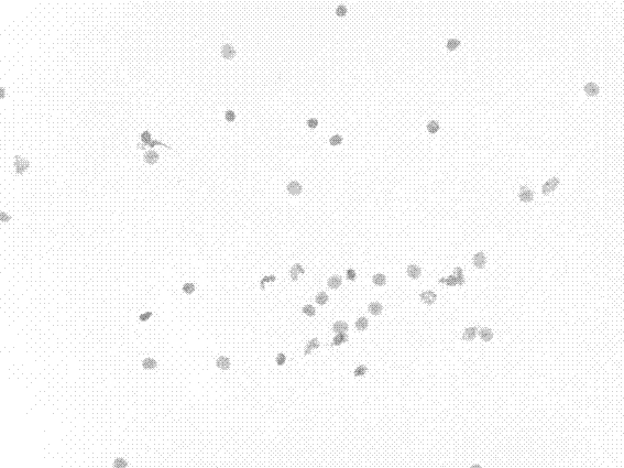
[0024] The pig liver cell smears were fixed in BS fixative solution for 25 minutes, hydrolyzed in 5N hydrochloric acid for 20 minutes, Thionin staining solution for 30 minutes, rinsed with tap water for 2 minutes between each step, and then rinsed with distilled water for 5 minutes, dehydrated with rapid gradient ethanol, and sealed. piece. BS fixative, 5N hydrochloric acid and Thionin staining solution all need to be pre-prepared in a 35°C incubator. The preparation process of Thionin dyeing solution is also carried out in a 35°C incubator. The preparation water needs to be preheated to 35°C in advance. The BS fixation process , Hydrochloric acid hydrolysis process and dyeing process are carried out in 35 ℃ constant temperature box. The stained pig liver smear under the microscope (20×) nuclei image as figure 1 shown.

Embodiment 2

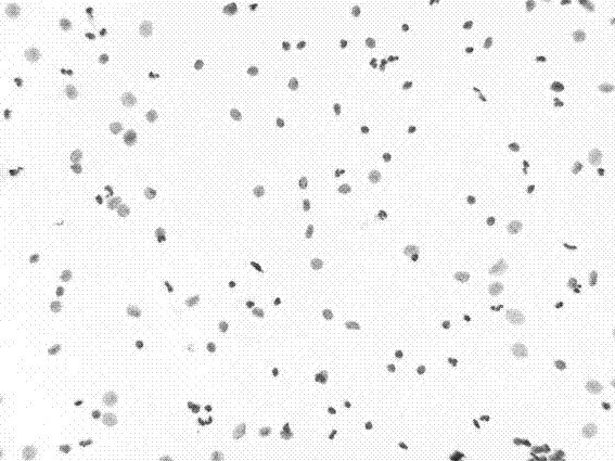
[0026] The cervical liquid-based cytology specimens were fixed in BS fixative solution for 30 minutes, hydrolyzed in 5N hydrochloric acid for 20 minutes, Thionin staining solution for 30 minutes, rinsed with tap water for 2 minutes between each step, and then rinsed with distilled water for 5 minutes, and dehydrated with rapid gradient ethanol , cover film. BS fixative solution, 5N hydrochloric acid and Thionin dye solution all need to be pre-prepared in a 40°C incubator. The preparation process of Thionin dye solution is also carried out in a 40°C incubator. The preparation water needs to be preheated to 40°C in advance. BS fixation process , Hydrochloric acid hydrolysis process and dyeing process are carried out in 40 ℃ incubator. The stained cervical liquid-based cytology specimens under the microscope (20×) nuclei images are as follows: figure 2 shown.

Embodiment 3

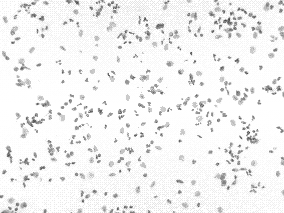
[0028] The cervical liquid-based cytology specimens were fixed in BS fixative solution for 30 minutes, hydrolyzed in 5N hydrochloric acid for 25 minutes, Thionin staining solution for 40 minutes, rinsed with tap water for 2 minutes between each step, and then rinsed with distilled water for 5 minutes, and dehydrated with rapid gradient ethanol , cover film. BS fixative solution, 5N hydrochloric acid and Thionin staining solution need to be pre-prepared in the 35°C automatic staining machine. The preparation process of Thionin staining solution is also carried out in the 35°C automatic staining machine. The preparation water needs to be preheated to 35°C in advance. BS The fixation process, the hydrochloric acid hydrolysis process and the dyeing process were all carried out in an automatic dyeing machine at 35°C. The stained cervical liquid-based cytology specimens under the microscope (20×) nuclei images are as follows: image 3 shown.


PUM


Abstract

The invention discloses a cell nucleus DNA (Deoxyribose Nucleic Acid) staining method. The cell nucleus DNA staining method is characterized by comprising the following steps: 1, fixation: sequentially placing sample slices into a BS fixing solution at the temperature of 30-40 DEG C to fix for 20-30 minutes, and rinsing; 2, hydrolyzation: completely fixing the sample slices, hydrolyzing in 5N hydrochloric acid at the temperature of 30-40 DEG C for 20-30 minutes, and rinsing; 3, staining: after the hydrolyzation is completed, staining in a Thionin staining solution at the temperature of 30-40 DEG C for 30-40 minutes, and rinsing; 4, after the staining is completed, dewatering by adopting fast gradient ethanol, and mounting. In conclusion, compared with other Feulgen staining methods, the cell nucleus DNA cell nucleus DNA has the obvious advantages of practicability, stability, efficiency, cost saving, repeatability, compatibility of automated equipment and the like and can meet the requirement of clinical DNA quantitative cytology on fast diagnosis.

Description

technical field [0001] The invention relates to the technical field of biological cell and tissue staining, in particular to a nuclear DNA staining method suitable for DNA quantitative cytology. Background technique [0002] Quantitative DNA cytology mainly stains the DNA in the nucleus, and then uses the image analysis system to measure the DNA content of the nucleus, so as to judge the physiological state and pathological changes of the cells. Like flow cytometry, it has been widely used in scientific research and clinical practice. At present, it has become a relatively advanced inspection technology for cancer and precancerous lesions in the world. [0003] At present, the DNA staining methods used in DNA quantitative cytology are mainly the traditional modified Feulgen method and the rapid Feulgen method. The traditional improved Feulgen method first fixes the cells in the specimen, and then hydrolyzes them with hydrochloric acid to break the glycosidic bond betwee...

Claims


Application Information

Patent Timeline
IPC IPC(8): G01N1/30
Inventor 唐剑黄荣祥
Owner MOTIC XIAMEN MEDICAL DIAGNOSTICS SYST
Why Eureka
Try Eureka