Check patentability & draft patents in minutes with Patsnap Eureka AI!

Method for assembling endoscopic imaging unit and endoscope

A technology of a camera unit and an assembling method, applied in the field of endoscopes, can solve the problems of the outer diameter of the tip portion becoming larger and larger, etc.

Inactive Publication Date: 2013-03-13
OLYMPUS CORP
View PDF7 Cites 3 Cited by
  • Summary
  • Abstract
  • Description
  • Claims
  • Application Information

AI Technical Summary

Problems solved by technology

[0007] However, the above-mentioned conventional example has the disadvantage that, for example, if the objective lens system includes a lens portion with a large outer diameter, it is necessary to also increase the size of the rear opening at the distal end, resulting in an increase in the outer diameter of the distal end.

Method used


Image

  • Method for assembling endoscopic imaging unit and endoscope
  • Method for assembling endoscopic imaging unit and endoscope
  • Method for assembling endoscopic imaging unit and endoscope

Examples


no. 1 Embodiment approach

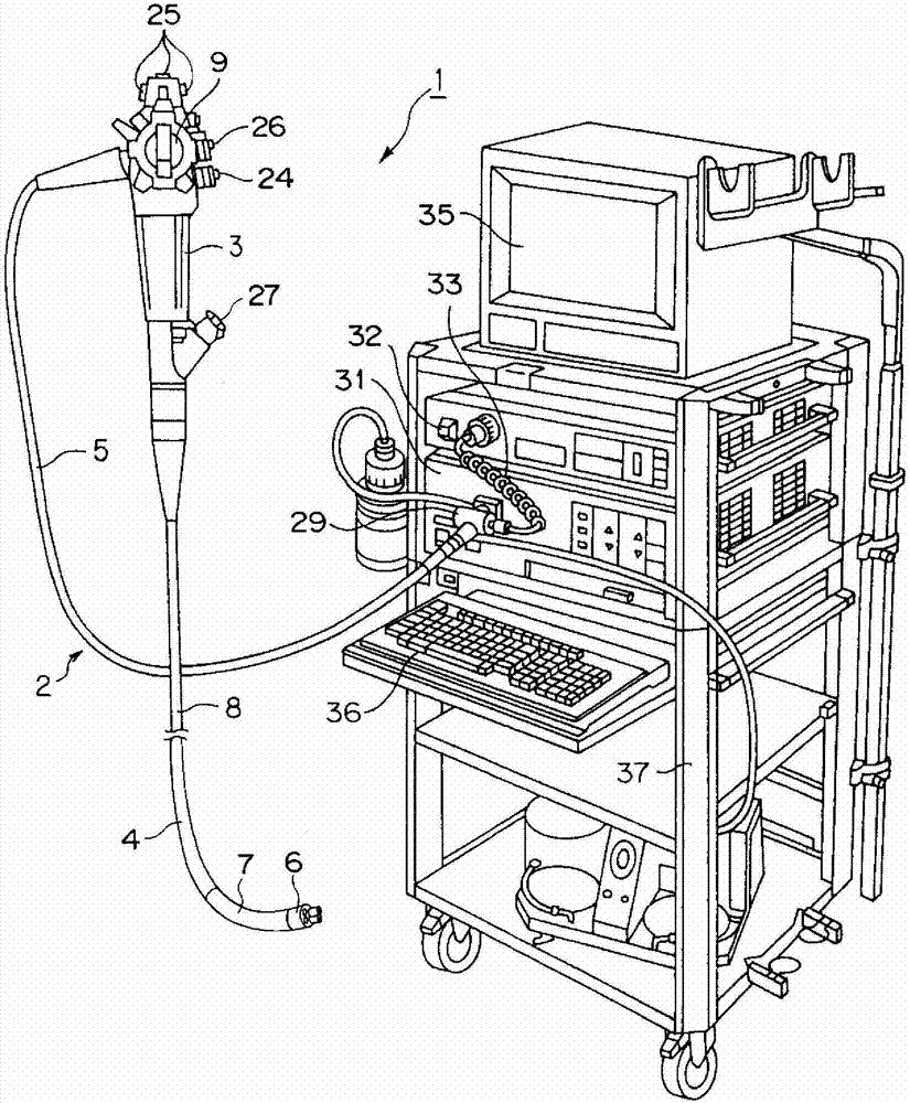
[0042] Such as figure 1 As shown, the endoscope apparatus 1 according to the first embodiment of the present invention has an endoscope 2 for performing endoscopic inspection. This endoscope 2 includes an operation part 3 for the operator to hold and operate, an elongated insertion part 4 formed at the front end of the operation part 3 and inserted into a body cavity, etc., and a base end extending from the side of the operation part 3. out of the Universal Cable 5.

[0043] In addition, the insertion part 4 includes a hard tip part 6 provided at the tip end, a freely bendable curved part 7 provided at the rear end of the tip part 6 , and a vertically long and flexible flexible part provided at the rear end of the curved part 7 . The flexible tube portion 8 and the bending portion 7 can be bent by a bending operation lever 9 provided on the operating portion 3 .

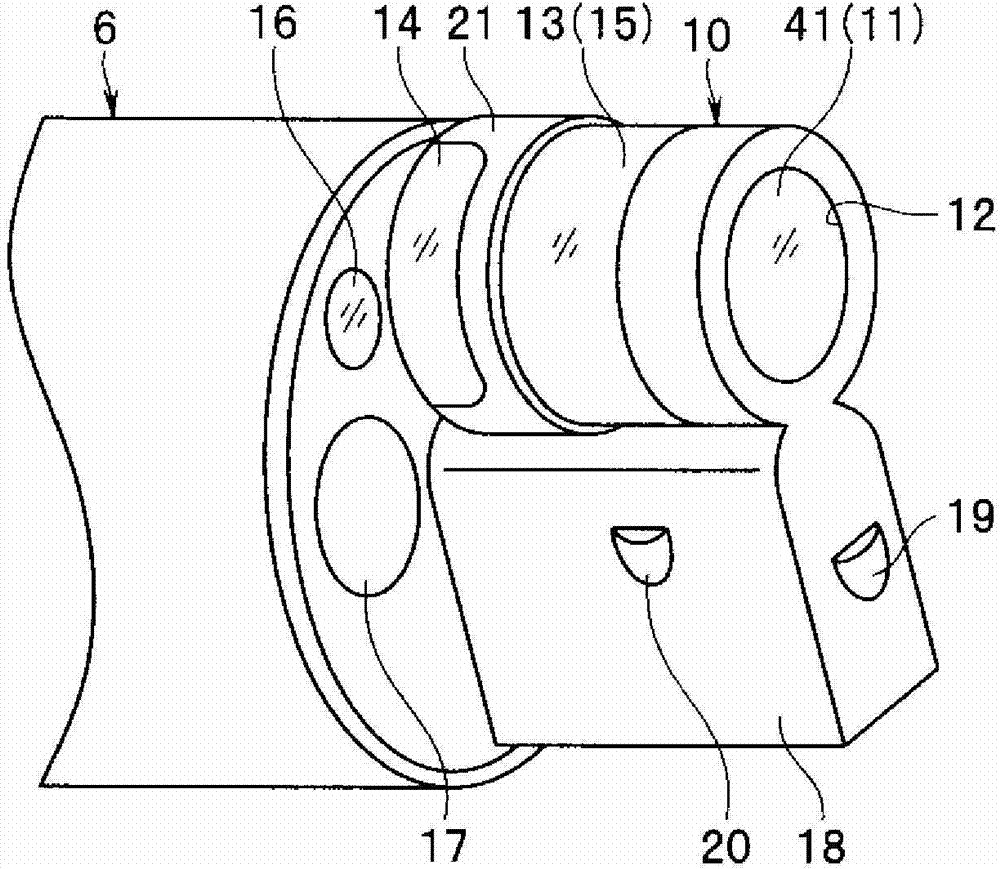
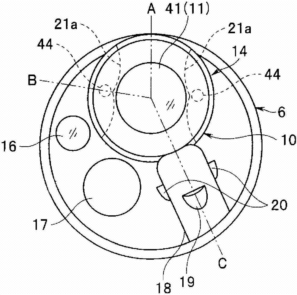
[0044] Additionally, if figure 2 As shown, a cylindrical distal end portion 10 is formed on the distal end por...

no. 2 Embodiment approach

[0124] Next, a second embodiment of the present invention will be described. Since this embodiment is similar to the first embodiment, different parts will be described. In the first embodiment, the condition of φ1>φa=φ2 (where φa=φ2 is set when the lens frame 56 is not provided as described above) is set. Assembled under different conditions. Specifically, assembly can be performed under the condition of φ2=φ1<φa=φ1′. Here, φ1 ′ represents the outer diameter of the rear end side of the lens frame 56 .

[0125] In addition, in this embodiment, the lens frame 56 separated from the front lens unit 51 is used before assembly, and the length L2 of the lens frame 56 in the optical axis direction is longer than that of the first embodiment.

[0126] However, this length L2 is set to be equal to or less than the opening length W of the side opening 64 so that the lens frame 56 can be inserted into the arrangement space 65 from the side opening 64 . In addition, in this embodiment...


PUM


Abstract

A method for assembling an endoscopic imaging unit provided with: a main tip body having a front opening, a side opening, a back opening, and a positioning space; a lens part comprising a tip lens with an outer diameter that substantially fits into the front opening; and an imaging part equipped with an image pick-up element that fits in the back opening and is disposed at the position where images are formed by a lens system such as the lens part. The method is provided with: an insertion step of inserting the lens part from the side opening into the positioning space; a fitting step of fitting the inserted lens part into the front opening; and an image pick-up element-fitting step of fitting the image pick-up element from behind the back opening into the back opening.

Description

technical field [0001] The present invention relates to an assembly method of an imaging unit of an endoscope in which an imaging unit is embedded in a distal end portion of an insertion portion, and an endoscope. Background technique [0002] In recent years, endoscopes in which an imaging unit is provided at the distal end of an insertion portion have been widely used in medical fields such as inspections in body cavities. [0003] When the insertion portion is inserted into a body cavity or the like, good insertion performance can be ensured by reducing the size of the imaging unit provided at the distal end portion of the insertion portion. Therefore, various methods of assembling the imaging unit fitted into the distal end portion of the insertion portion have been proposed. [0004] For example, in the conventional example of Japanese Unexamined Patent Publication No. 5-269081, an assembly method of an endoscope imaging unit is disclosed in which the imaging unit is i...

Claims


Application Information

Patent Timeline
IPC IPC(8): A61B1/04A61B1/00G02B23/24
CPCA61B1/00096G02B23/243H04N5/2253A61B1/00163Y10T29/49826H04N23/54
Inventor 本田一树
Owner OLYMPUS CORP
Features
  • R&D
  • Intellectual Property
  • Life Sciences
  • Materials
  • Tech Scout
  • Unparalleled Data Quality
  • Higher Quality Content
  • 60% Fewer Hallucinations
Learn More