Method for quantitative analysis for angiogenesis of living animals

An angiogenesis and quantitative analysis technology, applied in the field of quantitative angiogenesis in living animals, can solve the problems of not pointing out key indicators, not selecting a designated area, not analyzing the internal relationship of each parameter, etc.

Inactive Publication Date: 2013-09-11
XIDIAN UNIV
View PDF7 Cites 10 Cited by
  • Summary
  • Abstract
  • Description
  • Claims
  • Application Information

AI Technical Summary

Problems solved by technology

This article made a systematic analysis of imaging modes such as fluorescence microscopy, confocal and multiphoton microscopy, and electron microscopy, and pointed out that the biggest defect of these methods in clinical applications is that they cannot achieve in vivo imaging, and imaging The scope is too small to be suitable for the research of the overall goal, see: D.M.McDonald, P.L.Choyke, Imaging of angiogenesis: from microscope to clinic. Nature Medicine, 9(6):713-725(2003). At the same time, this two-dimensional The evaluation method has the following disadvantages: animals need to be sacrificed at different time points to obtain frozen sections of muscle tissue, and then to detect the presence of angiogenesis targets under a fluorescent microscope, so the analysis results are heavily dependent on the selection of slices. to the appropriate position, resulting in human misinterpretation, or even the slice does not select the designated area at all, which will lead to the possibility of a certain proportion of false negative results in the test
Different from the way of vascular casting, A. Arkudas et al. established a mouse arteriovenous ring model of in vitro cultured blood vessels. Since the blood v

Method used


Image

  • Method for quantitative analysis for angiogenesis of living animals
  • Method for quantitative analysis for angiogenesis of living animals
  • Method for quantitative analysis for angiogenesis of living animals

Examples


Embodiment Construction

[0051] Various details involved in the technical solution of the present invention will be described in detail below in conjunction with the accompanying drawings. It should be pointed out that the described embodiments are only intended to facilitate the understanding of the present invention, rather than limiting it in any way.

[0052] In this example, the angiogenesis in the lower limbs of mice was used as a quantitative object, but not limited thereto; under a stereomicroscope, the femoral artery and nerves on the inside of the lower limbs of the mice were dissected, and the femoral artery was ligated at the high position of the femoral artery; After adipose-derived mesenchymal stem cells were expanded and cultured to a sufficient number, the stem cells were injected into three adjacent positions within 1mm of the ligation site in the mouse lower limb ischemia model with a syringe.

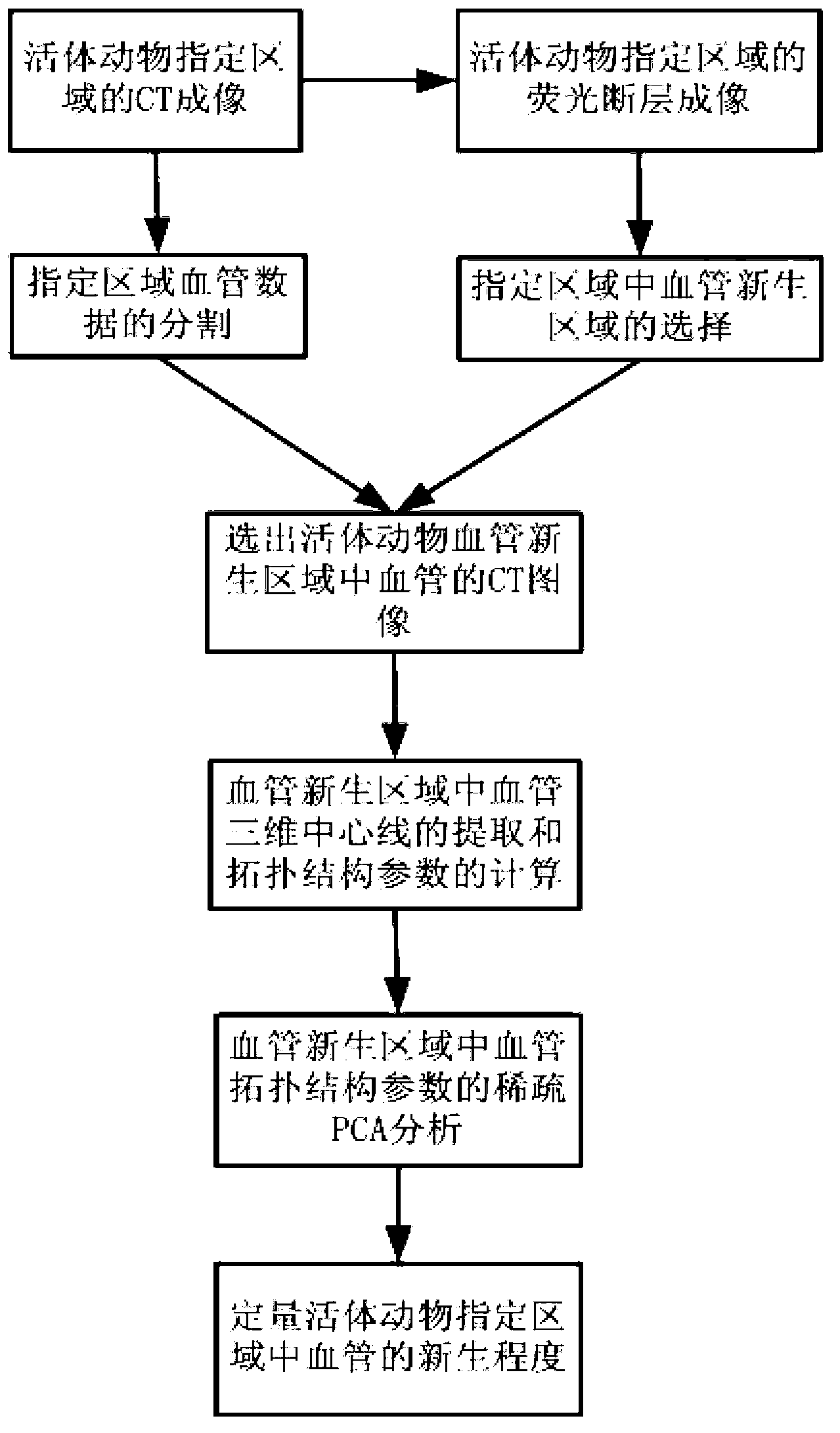
[0053] refer to figure 1 , the present invention is based on the quantitative method of ...


PUM


Abstract

Disclosed is a method for quantitative analysis for angiogenesis of living animals. The method includes steps of 1), CT (computed tomography) imaging in specified area of the living animal; 2), segmentation of vascular data; 3), fluorescence molecular tomography in the specified area of the living animal; 4), selection of CT images of the vascular in the specified area of the living animal; 5), analysis of vascular topology structure parameters by the aid of a sparse PCA (patient controlled analgesia) technology; 6), quantification of the angiogenesis. By the method, the quantification of the angiogenesis of the living animal can be achieved. The vascular in an angiogenesis area is selected by means of the fluorescence molecular tomography, precise quantification of the angiogenesis can be achieved based on the sparse PCA technology, and a quantitative method of the angiogenesis can be devised by the aid of the sparse PCA technology. During the quantification, different weighted values are distributed to each of the vascular topology structure parameters according to processing results of the sparse PCA technology, and contribution degrees of the different vascular topology structure parameters are distinguished.

Description

technical field [0001] The invention belongs to a method for quantitative analysis of angiogenesis in living animals. Specifically, the present invention belongs to the technical field of image processing, and relates to fluorescence tomography and CT imaging, especially a quantitative method for angiogenesis in living animals based on fluorescence tomography and CT imaging, which can be used for quantitative analysis of angiogenesis in living animals. Background technique [0002] At present, it is generally believed that promoting angiogenesis can treat ischemic diseases. Microscopic imaging of isolated tissue sections is often used to indirectly evaluate the degree of angiogenesis by observing the imaging of labeled probes. A review article published in Nature Medicine in 2003 summarized microscopic imaging studies of angiogenesis. This article made a systematic analysis of imaging modes such as fluorescence microscopy, confocal and multiphoton microscopy, and electron m...

Claims


Application Information

Patent Timeline
IPC IPC(8): A61B5/00A61B6/03
Inventor 屈晓超赵凤军易黄建冉超刘俊廷梁继民田捷
Owner XIDIAN UNIV
Why Eureka
Try Eureka