Ultrasonic angiography image analysis method and system thereof

A technology of image analysis and contrast-enhanced ultrasound, which is applied in image analysis, image enhancement, image data processing, etc., can solve the problems that the area to be punctured cannot be determined, the analysis results are meaningless, and there is no analysis involving speed, so as to achieve the benefit of clinical diagnosis. Effect

Active Publication Date: 2015-04-08
王本刚
View PDF0 Cites 11 Cited by
  • Summary
  • Abstract
  • Description
  • Claims
  • Application Information

AI Technical Summary

Problems solved by technology

[0008] 1) If the enhancement inside the area is uneven, such as necrotic areas, or honeycomb, etc., the analysis results of this area are meaningless;
[0009] 2) In hemangioma, etc., there are features such as ring enhancement, and the time displayed by the average intensity will not be able to show the ring enhancement in this area;
[0010] 3) In fields such as prostate puncture intervention, because the difference between regions is very small, it is impossible to determine the region to be punctured by using the region analysis method;
[0011] 4) The current regional analysis only involves the analysis of the strength, not the analysis of the change of the strength, that is, the analysis of the velocity; and

Method used


Image

  • Ultrasonic angiography image analysis method and system thereof
  • Ultrasonic angiography image analysis method and system thereof
  • Ultrasonic angiography image analysis method and system thereof

Examples


Embodiment Construction

[0060] In order to make the technical content disclosed in this application more detailed and complete, reference may be made to the drawings and the following various specific embodiments of the present invention, and the same symbols in the drawings represent the same or similar components. However, those skilled in the art should understand that the examples provided below are not intended to limit the scope of the present invention. In addition, the drawings are only for schematic illustration and are not drawn according to their original scale.

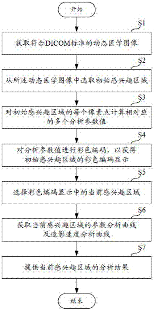
[0061] figure 1 A flow chart showing a method for analyzing a contrast-enhanced ultrasound image according to an embodiment of the present invention.

[0062] refer to figure 1 , in this contrast-enhanced ultrasound image analysis method, firstly, step S1 is executed to obtain a dynamic medical image conforming to the DICOM standard; then, step S2 is executed to select an initial region of interest from the dynamic medical imag...


PUM


Abstract

The invention provides an ultrasonic angiography image analysis method and system. The method comprises the following steps: obtaining a dynamic medical image meeting a DICOM (Digital Imaging and Communications in Medicine) standard; selecting an initial region of interest from the dynamic medical image; calculating a plurality of corresponding analysis parameter values of each pixel point of the initial region of interest, and carrying out colorful encoding on the analysis parameter values to obtain colorful codes of different parameters of the initial region of interest; selecting a current region of interest of the colorful codes; obtaining a parameter analysis curve of the current region of interest and a contrast speed analysis curve; providing an analysis result of the current region of interest. Compared with the prior art, the method can be used for carrying out the colorful encoding on the plurality of corresponding analysis parameter values of each pixel point, so that contrast parameters can be accurately quantified, and furthermore, accurate reference and differential diagnosis can be provided for doctors. The current region of interest can also be subjected to parameter analysis and speed analysis on the basis of the initial region of interest, and the clinical diagnosis can be facilitated.

Description

technical field [0001] The present invention relates to the technical field of contrast-enhanced ultrasound analysis, in particular to a method and system for analyzing contrast-enhanced ultrasound images. Background technique [0002] Ultrasonic instrument, also known as ultrasonic instrument, is a medical instrument developed based on the principle of ultrasound. It is an ultrasonic diagnostic device used in medical imaging systems in the fields of medical clinics, diagnosis, and hygiene. With the popularization of conventional gray-scale ultrasound and color Doppler ultrasound equipment, the role and value of these two ultrasound equipment in clinical diagnosis and treatment have become increasingly apparent, and they have become indispensable in clinical diagnosis. [0003] However, problems that cannot be solved by these two ultrasonic instruments often appear in clinical diagnosis, such as the display of low-speed blood flow and the detection of small blood vessels, be...

Claims


Application Information

Patent Timeline
Patent Type & Authority Applications(China)
IPC IPC(8): G06T7/00
CPCG06T7/0012G06T2207/10132G06T2207/30004
Inventor 王本刚
Owner 王本刚
Why Eureka
Try Eureka