Image-based ECG analysis method and apparatus

An electrical analysis and image technology, applied in the field of image-based electrocardiographic analysis methods and devices, can solve the problems of difficulty in data acquisition and analysis, inconvenience in electrocardiographic analysis, and the like

Inactive Publication Date: 2015-08-26
北京海思敏医疗技术有限公司
View PDF10 Cites 7 Cited by
  • Summary
  • Abstract
  • Description
  • Claims
  • Application Information

AI Technical Summary

Problems solved by technology

Usually there are certain requirements for the software architecture and installation environment, and the analysis and display of ECG data are completed together, which makes it difficult to realize long-term data collection and analysis. Inconvenient, such as doctors can only complete ECG analysis work in the office environment

Method used


Image

  • Image-based ECG analysis method and apparatus
  • Image-based ECG analysis method and apparatus
  • Image-based ECG analysis method and apparatus

Examples


Embodiment 1

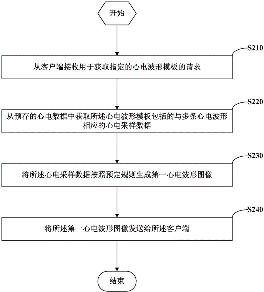
[0026] figure 2 It is a flow chart showing the image-based ECG analysis method according to Embodiment 1 of the present invention. The method may be performed, for example, on a server providing electrocardiographic data.

[0027] refer to figure 2 , in step S210, a request for acquiring a specified ECG waveform template is received from the client.

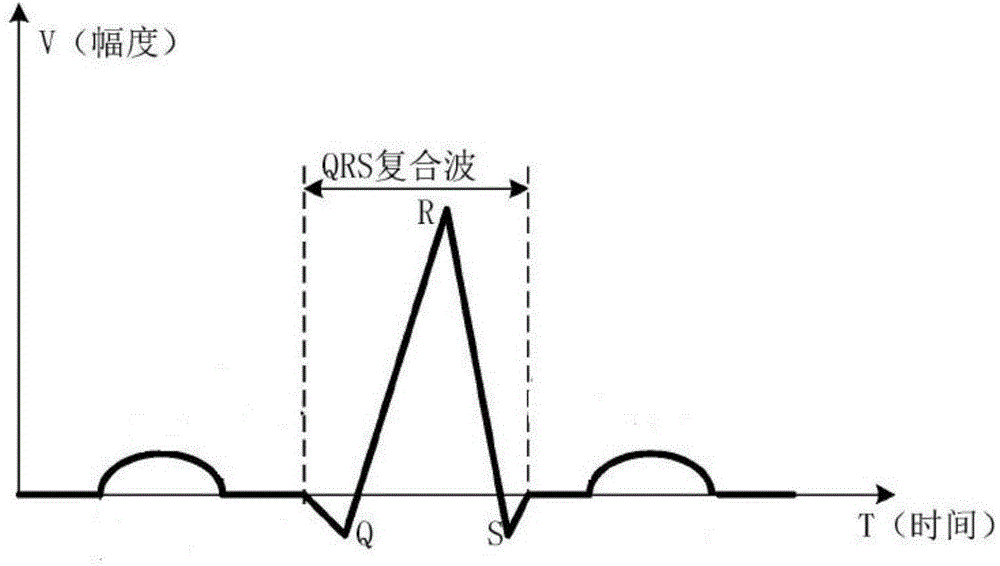
[0028] Here, the specified ECG waveform template can be, for example but not limited to, ventricular premature beat template, atrial premature beat template, supraventricular premature beat template, long pause template, atrial flutter template, atrial fibrillation template, ventricular flutter template or ventricular fibrillation template template.

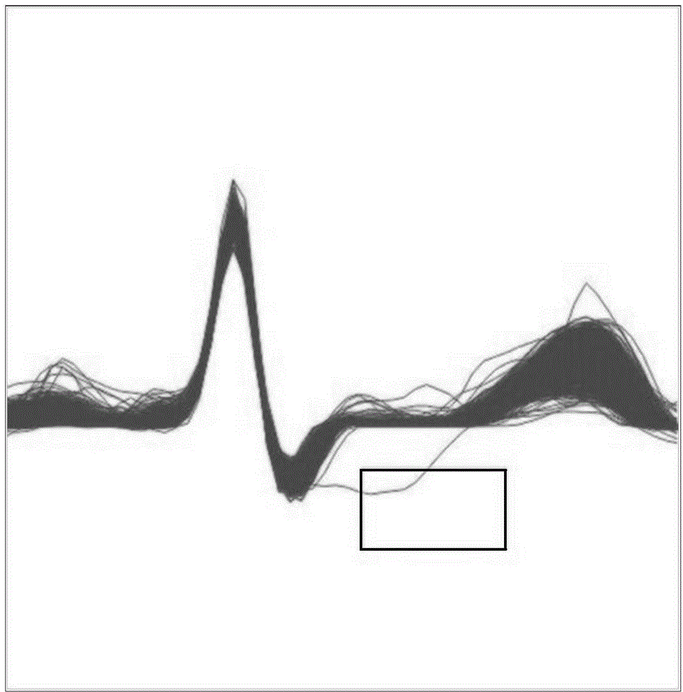
[0029] In step S220, ECG sampling data corresponding to a plurality of ECG waveforms included in the ECG waveform template is obtained from prestored ECG data.

[0030] Specifically, for example, the server pre-stores the ECG data collected by the ECG acquisition device. After...

Embodiment 2

[0046] Figure 4 It is a flow chart showing the image-based ECG analysis method according to Embodiment 2 of the present invention. The method can be performed, for example, on a client.

[0047] refer to Figure 4 , in step S410, receiving a user instruction for acquiring a specified ECG waveform template.

[0048] Here, for example, a graphical user interface can be provided on the client, on which a plurality of icons representing ECG waveform templates can be displayed, and the user can send a user instruction for obtaining a specified ECG waveform template to the client by clicking on an icon. It should be noted that the specified ECG waveform template can be, for example but not limited to, a premature ventricular contraction template, atrial premature contraction template, supraventricular premature contraction template, long intermittent template, atrial flutter template, atrial fibrillation template, ventricular flutter template or ventricular fibrillation template...

Embodiment 3

[0056] Figure 5 It is a logic block diagram showing the image-based electrocardiogram analysis device according to the third embodiment of the present invention. can be used to execute as figure 2 The method steps of the illustrated embodiment.

[0057] refer to Figure 5 , the image-based ECG analysis device includes an ECG waveform template request receiving module 510, an ECG sampling data acquisition module 520, a first ECG waveform image generation module 530 and a first ECG waveform image sending module 540, wherein:

[0058] The ECG waveform template request receiving module 510 is configured to receive a request for obtaining a specified ECG waveform template from a client.

[0059] The electrocardiographic sampling data acquisition module 520 is configured to acquire electrocardiographic sampling data corresponding to a plurality of electrocardiographic waveforms included in the electrocardiographic waveform template from prestored electrocardiographic data.

[...


PUM


Abstract

Embodiments of the present invention provide an image-based ECG analysis method and apparatus. The method comprises: receiving from a client a request for acquiring a designated ECG waveform template; acquiring from prestored ECG data ECG sampling data corresponding to a plurality of ECG waveforms included in the ECG waveform template; generating the ECG sampling data into a first ECG waveform image according to a predetermined rule; and sending the first ECG waveform image to the client. The image-based ECG analysis method and apparatus provided in the embodiments of the present invention can separate display of the ECG data from analysis of the ECG data, implement long-term data collection and analysis, and hence achieve more convenient and flexible ECG analysis.

Description

technical field [0001] The invention relates to the technical field of electrocardiogram analysis, in particular to an image-based electrocardiogram analysis method and device. Background technique [0002] Usually, template analysis is a common method for dynamic ECG analysis. It can help doctors quickly find out abnormal heartbeats from hundreds of thousands of heartbeats, thereby assisting doctors in diagnosing patients. Most of the existing ambulatory electrocardiogram analysis systems run on stand-alone systems or are software based on C / S architecture. Usually there are certain requirements for the software architecture and installation environment, and the analysis and display of ECG data are completed together, which makes it difficult to realize long-term data collection and analysis. Inconvenient, for example, doctors can only complete ECG analysis work in the office environment. Contents of the invention [0003] The purpose of the embodiments of the present ...

Claims


Application Information

Patent Timeline
Patent Type & Authority Applications(China)
IPC IPC(8): G06F19/00
Inventor 邵永安王锋侯桂文
Owner 北京海思敏医疗技术有限公司
Why Eureka
Try Eureka