Optical coherent image segmentation method for retina

An optical coherence and image segmentation technology, applied in image analysis, image enhancement, image data processing and other directions, can solve the problems of time-consuming, insufficient practicability, slow speed, etc., and achieve accurate edge detection results, strong practicability, and computing speed. quick effect

Inactive Publication Date: 2016-08-24
HUNAN SCI & TECH RES & DEV CENT
View PDF3 Cites 11 Cited by
  • Summary
  • Abstract
  • Description
  • Claims
  • Application Information

AI Technical Summary

Problems solved by technology

Mujat et al. used the deformation spline method to segment the retinal optic nerve layer. It needs to place the deformation spline near the initial contour, which is usually time-consuming.
Chiu pointed out that most of the methods reported in the literature for 2D and 3D OCT image segmentation are slow, which makes them less practical in clinical practice.

Method used


Image

  • Optical coherent image segmentation method for retina
  • Optical coherent image segmentation method for retina
  • Optical coherent image segmentation method for retina

Examples


Embodiment Construction

[0018] 1. Data source

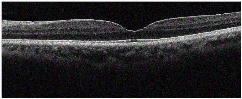
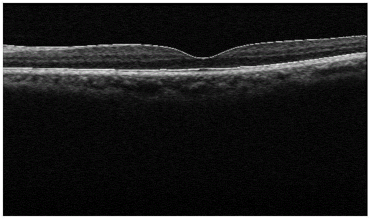
[0019] Using the optical coherence tomography method, we collected a total of 311 macular images from 34 patients, and the resolution of the original image was 2000 (depth direction) * 2048 (width direction).

[0020] 2. Macular image segmentation method based on multi-resolution and level set

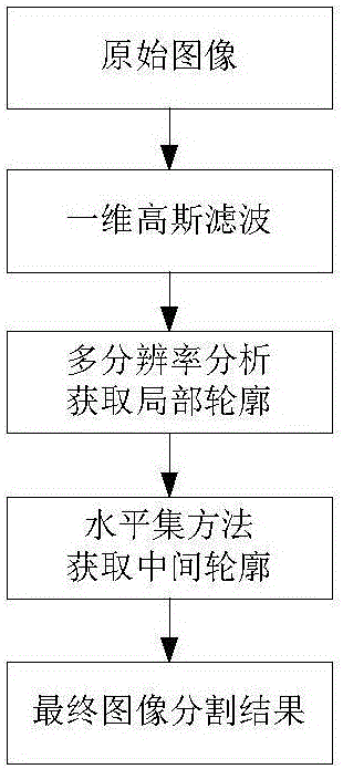
[0021] In order to assist the medical macular thickness measurement, it is necessary to obtain a clear outline of the macular image. The present invention designs a new macular image segmentation method based on multi-resolution and level sets. First, the original image is filtered by one-dimensional Gaussian filtering. Then use the multi-resolution method to obtain the initial local contour of the image, and finally use the level set method to quickly obtain the middle contour of the macular image to obtain the final image segmentation result. The process of the inventive method is as figure 1 shown.

[0022] (1) Gaussian filter

[0023] Gaussian filtering ...


PUM


Abstract

The invention discloses an optical coherent image segmentation method for a retina. Filtering is carried out on a retina macula lutea image line by line by means of Gaussian filter; an initial local contour of the image is obtained by using a multi-resolution-ratio method; and then a middle contour of the retina macula lutea image is obtained rapidly by using a level set method to obtain a final image segmentation result. According to the invention, the edge detection result is accurate and the calculation speed is fast. Retina segmentation can be completed without an initial seed point. The method can be applied to the clinic practice conveniently; and the practicability is high.

Description

technical field [0001] The invention relates to a retinal optical coherent image segmentation method. Background technique [0002] In the medical field, retinal macular thickness can be used to quantify diseases such as diabetic macular edema and age-related macular degeneration. Clinically, optical coherence tomography is usually used to obtain macular images. However, the existing macular image segmentation methods are slow in operation, which hinders their clinical application. Early OCT retinal image segmentation methods are mainly based on gray level thresholding and gray level variation, which are sensitive to noise and time-consuming. Koozekanani et al proposed a Markov random field (MRF) method to extract the inner and outer edges of the retina, and the robustness of this autoregressive model is superior to those based on gray thresholding. But it needs reliable initial seed points to complete the segmentation of pathological retina. Mujat et al. used the deforma...

Claims


Application Information

Patent Timeline
Patent Type & Authority Applications(China)
IPC IPC(8): G06T7/00
CPCG06T2207/10101G06T2207/20024G06T2207/30041
Inventor 张天桥黎日昌罗文
Owner HUNAN SCI & TECH RES & DEV CENT
Why Eureka
Try Eureka