OCT (Optical Coherence Tomography) image layer segmentation method based on neural network and constraint graph search algorithm

A neural network and search algorithm technology, applied in the field of medical image processing algorithms, can solve the problems of low contrast in the retinal layer of the image, large changes in the structure of the retinal layer, and blurred boundaries.

Active Publication Date: 2017-11-24
SUZHOU UNIV
View PDF4 Cites 20 Cited by
  • Summary
  • Abstract
  • Description
  • Claims
  • Application Information

AI Technical Summary

Problems solved by technology

This results in low contrast and blurred boundaries between retinal layers in OCT images, and also highly variable retinal layer structure
Therefore, layer segmentation us

Method used


Image

  • OCT (Optical Coherence Tomography) image layer segmentation method based on neural network and constraint graph search algorithm
  • OCT (Optical Coherence Tomography) image layer segmentation method based on neural network and constraint graph search algorithm
  • OCT (Optical Coherence Tomography) image layer segmentation method based on neural network and constraint graph search algorithm

Examples


Embodiment 1

[0070] This embodiment is based on the OCT image segmentation method of neural network and constraint graph search algorithm, including:

[0071] Obtain OCT image feature training neural network classifier;

[0072] Obtain the final SF1 using a multi-resolution graph search algorithm;

[0073] Extract 24 features of the OCT image, use a neural network classifier to classify and mark the OCT image into 8 regions, and find the upper surface of the marked region from top to bottom as the initial boundaries S1, S2,..., S8 of the 8 retinal regions;

[0074] According to the initial boundaries S2 to S8, use the constraint graph search algorithm to find the precise SF2 to SF8 in sequence;

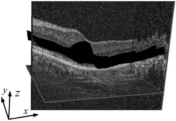
[0075] Neovascularization and effusion were segmented between SF7 and SF8.

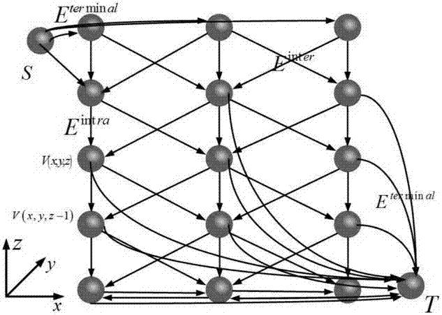
[0076] Among them, the constraint graph search algorithm specifically includes:

[0077] Step 1: The OCT image to be segmented uses the OCT image after anisotropic filtering and respond according to the multiscale br...

Embodiment 2

[0095] This embodiment is based on the OCT image segmentation method of neural network and constraint graph search algorithm, on the basis of embodiment 1, obtains OCT image feature training neural network classifier specifically includes:

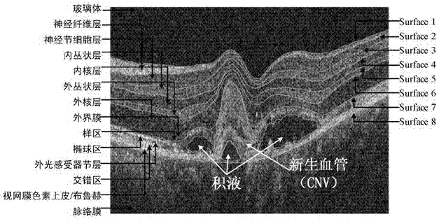
[0096] The retinal OCT images of choroidal neovascular retinopathy were divided into 8 regions; the divisions and the upper surface labels corresponding to each layer structure are as follows: region 1: nerve fiber layer SF1; region 2: ganglion cell layer SF2; region 3: inner plexus SF3; Region 4: Inner core layer SF4; Region 5: Outer plexiform layer SF5; Region 6: Outer nuclear layer + external membrane + sample region SF6; Region 7: Ellipsoid region + outer photoreceptor nodal layer + staggered region RPE / Bruch SF7; area 8: vitreous + choroid SF8.

[0097] Extract 24 features of the OCT image, the specific method is as follows:

[0098]Step 1: Let the upper surface of area 1 in the OCT image be SF1. Find the scan from SF1 to the bottom...

Embodiment 3

[0114] This embodiment is based on the OCT image segmentation method of the neural network and the constraint graph search algorithm. On the basis of Embodiment 1 and Embodiment 2, the neural network classifier used to obtain the OCT image features specifically includes:

[0115] Extract 24 features of the OCT image, the specific method is as follows:

[0116] Step 1: Set the coordinate vector of the pixel in the OCT image OCT images using a Gaussian filter Filtering is performed, and then the initial shape of Surface1 is found using the canny edge detection algorithm; the precise shape SF1 of Surface1 is based on the initial shape and the multi-resolution graph search algorithm. Find the scan from SF1 to the bottom, and calculate the distance from the pixel below SF1 to SF1 as a distance feature; the horizontal axis coordinate x and vertical axis coordinate y of the OCT image are used as the other two features, where the surface SF of the retinal layer can be determined by...


PUM


Abstract

The invention relates to an OCT (Optical Coherence Tomography) image layer segmentation method based on a neural network and a constraint graph search algorithm, and is designed for accurately segmenting a retina layer and new vessels. The OCT image layer segmentation method based on the neural network and the constraint graph search algorithm comprises the following steps that: obtaining OCT image features to train a neural network classifier; obtaining a final SF1 through a multi-resolution map search algorithm SF1; extracting 24 features of an OCT image, and using the neural network classifier to find initial surfaces S1, S2,...S8; according to initial boundaries S2 to S8, using the constraint graph search algorithm to find accurate SF2 to SF8 in sequence; and segmenting the new vessels and hydrops between SF7 and SF8. The OCT image layer segmentation method based on the neural network and the constraint graph search algorithm has the advantages of being simple in operation and accurate in detection results. The existing problems of low identification rate, poor segmentation effect and the like of a lesion OCT image segmentation algorithm.

Description

technical field [0001] The invention belongs to the field of medical image processing algorithms, in particular to an OCT image layer segmentation method based on a neural network and a constraint graph search algorithm. Background technique [0002] Age-related macular degeneration (also known as age-related macular degeneration, AMD) is a degenerative disease of the macula. leading cause of blindness. AMD often occurs in both eyes, with abnormal depigmentation or hyperpigmentation changes in the pigment epithelium (RPE), geographic atrophy of the pigment epithelium and choroidal capillaries, choroidal neovascularization (CNV) formation, and macular exudation. Therefore, the quantitative analysis of age-related macular degeneration retinopathy has very important significance in the research of retina. [0003] Optical coherence tomography (OCT) can quickly and non-invasively clearly display the lesions of each layer of the retina, and is the only non-invasive examination ...

Claims


Application Information

Patent Timeline
IPC IPC(8): G06T7/10G06T7/187G06N3/08
CPCG06T7/10G06T7/187G06T2207/10101G06T2207/20081G06T2207/20084G06T2207/30041G06T2207/30101
Inventor 向德辉
Owner SUZHOU UNIV
Why Eureka
Try Eureka