
Slit-lamp microscope for 3D examination of eyelid plate gland

A slit lamp microscope and eyelid plate technology, applied in the field of microscopy, can solve the problems of light sources from other bands, unable to meet the doctor's observation needs, and the image is not clear enough

Active Publication Date: 2018-01-16
杭州爱视界医疗器械有限公司
View PDF24 Cites 3 Cited by
  • Summary
  • Abstract
  • Description
  • Claims
  • Application Information

AI Technical Summary

Problems solved by technology

[0003] The current slit lamp microscope usually adopts the method of adding an infrared camera to collect the weak infrared light in the compound light source of the slit lamp reflected by the human meibomian glands, and finally form an image to check the condition of the meibomian gland patients, because the infrared light is extremely It is dark and weak, and it is also mixed with a large number of light sources of other wavelength bands, so the image obtained in this way is not clear enough and the image is flat, which cannot meet the doctor's observation needs. At present, there is no good way to solve the above problems. This problem needs to be solved

Method used


Image

  • Slit-lamp microscope for 3D examination of eyelid plate gland
  • Slit-lamp microscope for 3D examination of eyelid plate gland
  • Slit-lamp microscope for 3D examination of eyelid plate gland

Examples


Embodiment Construction

[0022] Hereinafter, the present invention will be further described through specific embodiments in conjunction with the drawings.

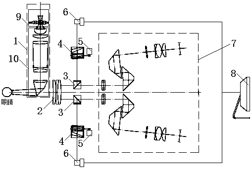
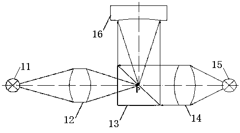
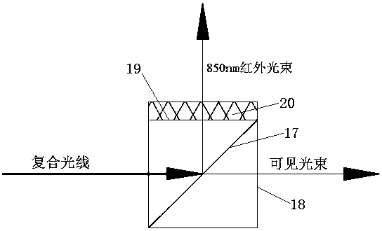
[0023] Such as Figure 1 to Figure 4 As shown, a slit lamp microscope capable of 3D inspection of the eyelid meibomian glands includes an illumination system 1, a large objective lens 2, a dichroic filter prism 3, a taper scanning objective lens group 4, a motor 5, a CCD 6, a vision system 7 and a computer 8. The taper scanning objective lens group 4 is connected to the motor 5, the CCD 6 is connected to the computer 8, the illumination system 1, the large objective lens 2, the dichroic filter prism 3, the taper scanning objective lens group 4, and the CCD 6 are arranged in sequence along the light incident direction. The illumination system 1, the large objective lens 2, the dichroic filter prism 3, and the visual system 7 are arranged in sequence along the light incident direction.

[0024] Specifically, the illumination system 1 includes a two-col...


PUM


Abstract

The invention discloses a slit-lamp microscope for 3D examination of an eyelid plate gland. The slit-lamp microscope comprises an illumination system, a large objective lens, a light splitting and color filtering prism, a taper scanning objective lens set, a motor, a CCD, a visual system and a computer, wherein the taper scanning objective lens set is connected with the motor, the CCD is connectedwith the computer, the illumination system, the large objective lens, the light splitting and color filtering prism, the taper scanning objective lens set and the CCD are sequentially arranged in thelight incidence direction, and the illumination system, the large objective lens, the light splitting and color filtering prism and the visual system are sequentially arranged in the light incidencedirection. The slit-lamp microscope for 3D examination of the eyelid plate gland adopts a double-color compound-point light source structure, the light splitting and color filtering prism, the taper scanning objective lens set and the like, and can produce high-definition 3D infrared images and provide a basis for a doctor to examine the eyelid plate gland of a diseased patient.

Description

Technical field [0001] The invention relates to the technical field of microscopes, in particular to a slit lamp microscope capable of 3D inspection of the eyelid glands. Background technique [0002] Slit lamp microscope is an important instrument indispensable for ophthalmological examinations. Usually, the slit lamp microscope consists of an illumination system and a binocular microscope. It not only enables the superficial lesions to be observed very clearly, but also adjusts the focus and the width of the light source. The "optical section" makes the lesions of deep tissues clearly visible. [0003] The current slit lamp microscope usually adopts the method of adding an infrared camera to collect the weak infrared light from the slit lamp compound light source reflected by the human eye meibomian glands, and finally image the image to check the condition of the eyelid meibomian glands, because the infrared light is extremely It is dark and weak, and also contains a large numb...

Claims


Application Information

Patent Timeline
Patent Type & Authority Applications(China)
IPC IPC(8): A61B3/135
CPCY02A40/81
Inventor 程得集
Owner 杭州爱视界医疗器械有限公司
Learn More