Angiography image blood vessel segmentation method and system

A technology in contrast images and images, which is applied in the field of image processing, can solve the problems of increasing the workload of doctors and cumbersome interactive operations, etc., and achieve the effect of clear blood vessel boundaries, obvious details, and high efficiency

Active Publication Date: 2018-06-22
BEIJING INSTITUTE OF TECHNOLOGYGY
View PDF6 Cites 23 Cited by
  • Summary
  • Abstract
  • Description
  • Claims
  • Application Information

AI Technical Summary

Problems solved by technology

[0006] Model-based segmentation method, the advantage of this method is that the accuracy is relatively high, and the background noise can be avoided by initially selecting the seed point, but the disadvantage is that it requires interaction
Because there are a large number of artifact structures in the background area of ​​contrast images, the most widely used segm

Method used


Image

  • Angiography image blood vessel segmentation method and system
  • Angiography image blood vessel segmentation method and system
  • Angiography image blood vessel segmentation method and system

Examples


Embodiment Construction

[0049] The specific implementation manners of the present invention will be further described in detail below in conjunction with the accompanying drawings and embodiments. The following examples are used to illustrate the present invention, but are not intended to limit the scope of the present invention.

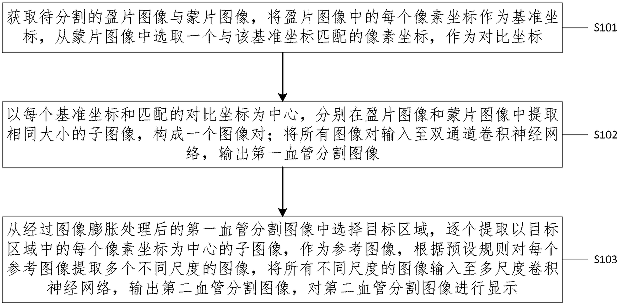
[0050] combine figure 1 , is a schematic flowchart of a method for segmenting blood vessels in contrast images provided by an embodiment of the present invention. This embodiment describes a method for multi-scale display of blood vessels in contrast images based on the present invention. It should be noted that this The multi-scale described in the various embodiments of the invention may be the caliber of the blood vessel, or other parameters capable of distinguishing different types of blood vessels.

[0051] The method includes:

[0052] 101. Acquire the image to be segmented and the mask image, use each pixel coordinate in the image as a reference coordinate, and se...


PUM


Abstract

The invention provides an angiography image blood vessel segmentation method and an angiography image blood vessel segmentation system. The angiography image blood vessel segmentation method includesthe steps of taking each pixel coordinate of a live image as a reference coordinate, selecting one pixel coordinate matched with the reference coordinate from a mask image and taking the pixel coordinate as a contrast coordinate; extracting sub images in the same size from the live image and the mask image respectively to form an image pair; inputting all the images to a dual-passage convolutionalneural network to output a first blood vessel segmentation image; extracting multiple images in different size from each reference image according to preset rules, inputting all the images in different size to a multi-scale convolutional neural network to output a second blood vessel segmentation image, and finally displaying the second blood vessel segmentation image. As the dual-passage convolutional neural network and the multi-scale convolutional neural network are utilized for achieving twice classification, blood vessels of the images displayed finally have defined boundaries and cleardetails, and during the whole process, full-automatic interaction is achieved with high efficiency.

Description

technical field [0001] The present invention relates to the technical field of image processing, and more particularly, to a method and system for segmenting blood vessels in contrast images. Background technique [0002] Today, coronary artery disease has become a major threat to human health. In the diagnosis and treatment of coronary heart disease, cardiovascular angiography images have become the gold standard for diagnosis. The computer-aided diagnosis and treatment system based on angiography images can help doctors diagnose the disease and formulate treatment plans, which has very important research significance. In the computer-aided diagnosis system of contrast images, blood vessel segmentation technology is the key technology, and it is the basis of center line extraction, radius measurement, three-dimensional reconstruction and other technologies. [0003] Contrast images have disadvantages such as low contrast, uneven distribution of contrast agents, and severe ...

Claims


Application Information

Patent Timeline
IPC IPC(8): G06T7/10G06T7/30
CPCG06T7/10G06T7/30G06T2207/20081G06T2207/20084G06T2207/30101
Inventor 杨健艾丹妮杨思远王涌天丛伟建蒋玉蓉
Owner BEIJING INSTITUTE OF TECHNOLOGYGY
Why Eureka
Try Eureka