Method for detecting a dental object

An object, dental technology, applied in the field of capturing dental objects, to achieve the effect of shortening the image acquisition time

Active Publication Date: 2018-06-29
SIRONA DENTAL SYSTEMS
View PDF7 Cites 4 Cited by
  • Summary
  • Abstract
  • Description
  • Claims
  • Application Information

AI Technical Summary

Problems solved by technology

[0006] A disadvantage of this method is that the sampling of isotropic volume datasets is associated with significantly longer image acquisition times compared to individual MRT cross-sectional images
Furthermore, the image quality of volumetric datasets is degraded due to subsequent image manipulations, precisely due to interpolation losses

Method used


Image

  • Method for detecting a dental object

Examples


Embodiment Construction

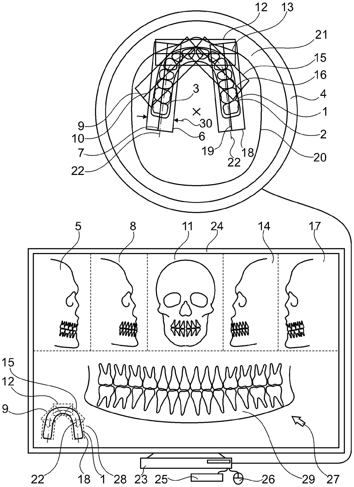
[0090] figure 1 A diagram is shown illustrating the current method for capturing at least a portion of a dental object 1 , in particular a maxilla 2 and / or a mandible 3 , by means of an MRT device 4 .

[0091] In this case, the first MRT segment image 5 of the first segmented volume 6 is acquired with the center plane 7; the second MRT segment image 8 of the second segmented volume 9 is acquired with the center plane 10; the second MRT segment image 8 is acquired with the center plane 13 A third MRT slice image 11 of the third segmented volume region 12; a fourth MRT slice image 14 of the fourth segmented volume region 15 is acquired with the central plane 16; and a fourth MRT segmented image 14 of the fifth segmented volume region 18 is acquired with the central plane 19 Five MRT fragment images 17 . In this context, the MRT segment images 5 , 8 , 11 , 14 and 17 form segment volumes 6 , 9 , 12 , 18 which overlap to a certain extent. To capture the dental object 1 , the pati...


PUM


Abstract

Disclosed herein is a method for capturing a dental object (1) with an object volume, in particular, at least one part of a maxilla (2) and/or a mandible (3), with the use of an MRT apparatus (4). A plurality of MRT segment images (5, 8, 11, 14, 17), which depict the defined segmented volume regions (6, 9, 12, 15, 18) are acquired by an MRT apparatus (4) within a measurement volume (21) of the MRTapparatus (4). The segmented volume regions (6, 9, 12, 15, 18) overlap no more than partially. In this case a target surface (22) is defined or has already been defined. Then a two dimensional composite image (29), which corresponds to a two dimensional aggregate image through the target surface (22) within the object volume of the dental object (1), is generated in a computer-aided manner from the individual MRT segment images (5, 8, 11, 14, 17).

Description

technical field [0001] The invention relates to a method for capturing a dental object having an object volume, specifically at least a part of the maxilla and / or the mandible, by means of an MRT device, wherein the MRT device A plurality of MRT slice images depicting defined segmented volume regions are acquired within the measurement volume of the device, wherein the segmented volume regions at most partially overlap. Background technique [0002] The prior art discloses several methods for generating MRT images of dental objects or parts thereof, such as maxilla / mandible. [0003] According to a known method, conventional MRT equipment is used to obtain individual MRT cross-sectional images of cuts such as the jaw arch. If the aim is to acquire the entire jaw arch, for example five individual MRT cross-sectional images will be necessary. [0004] One disadvantage of this method lies in the fact that manual planning of individual MRT cross-sectional images is associated ...

Claims


Application Information

Patent Timeline
Patent Type & Authority Applications(China)
IPC IPC(8): G01R33/483A61B5/055
CPCA61B5/055G01R33/483G01R33/4833G01R33/4835G01R33/4836A61C9/0046A61B5/7425A61B5/4547A61B5/7278A61B5/742A61B6/14A61B2576/02G06T5/50G06T7/0014G06T11/008G06T2207/10088G06T2207/10116G06T2207/30036
Inventor C.阿布凯T.布劳恩J.保罗V.拉舍
Owner SIRONA DENTAL SYSTEMS
Why Eureka
Try Eureka