3D lung nodule generation method, device and electronic device

A pulmonary nodule, 3D technology, applied in the field of image recognition, can solve problems such as general effect, inability to generate 3D pulmonary nodules, and difficulty in deep learning networks.

Inactive Publication Date: 2019-01-04
BEIJING PEREDOC TECH CO LTD
View PDF9 Cites 9 Cited by
  • Summary
  • Abstract
  • Description
  • Claims
  • Application Information

AI Technical Summary

Problems solved by technology

[0004] However, the existing labeled high-quality medical CT image data is still relatively lacking, which makes it difficult for the deep learning network to be fully trained, and there is a situation of overfitting, that is, the de

Method used


Image

  • 3D lung nodule generation method, device and electronic device
  • 3D lung nodule generation method, device and electronic device
  • 3D lung nodule generation method, device and electronic device

Examples


Embodiment 1

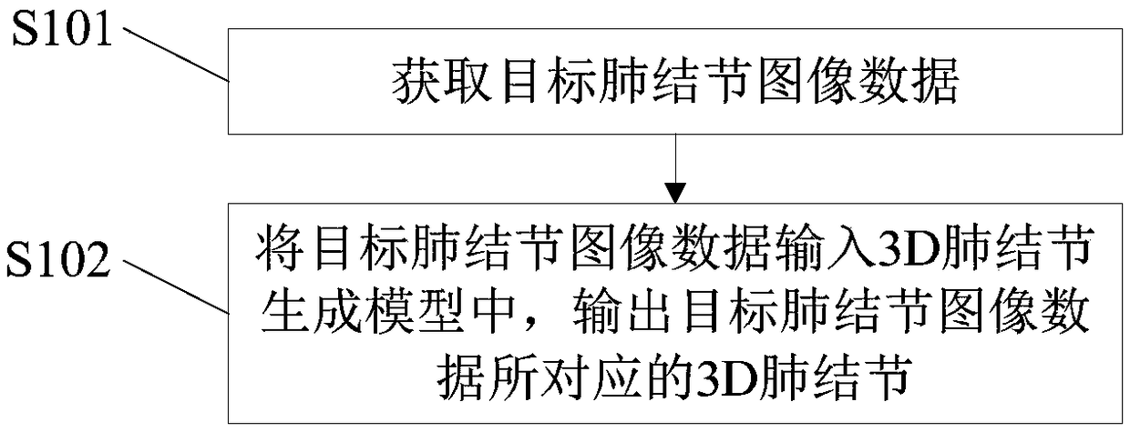
[0055] The embodiment of the present invention provides a method for generating 3D pulmonary nodules, see figure 1 As shown, the method includes the following steps:

[0056] S101: Acquire target pulmonary nodule image data.

[0057] S102: Input the target pulmonary nodule image data into the 3D pulmonary nodule generating model, and output the 3D pulmonary nodule corresponding to the target pulmonary nodule image data.

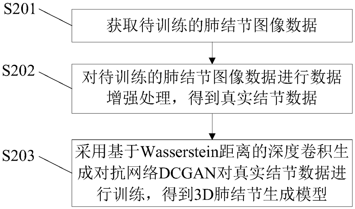
[0058] Among them, the 3D pulmonary nodule generation model is obtained by training the real nodule data through the deep convolution generation confrontation network DCGAN based on the Wasserstein distance; the real nodule data is obtained by data enhancement processing of the pulmonary nodule image data to be trained.

[0059] The following is a detailed description of the establishment process of the 3D pulmonary nodule generation model, see figure 2 Shown:

[0060] S201: Obtain image data of pulmonary nodules to be trained.

[0061] First read the me...

Embodiment 2

[0086] The embodiment of the present invention also provides a 3D pulmonary nodule generation device, see Figure 4 As shown, the device includes: a first data acquisition module 41 and a 3D pulmonary nodule generation module 42 .

[0087] Wherein, the first data acquisition module 41 is used to acquire target pulmonary nodule image data; the 3D pulmonary nodule generation module 42 is used to input the target pulmonary nodule image data into the 3D pulmonary nodule generation model and output the target pulmonary nodule 3D lung nodules corresponding to the image data.

[0088] The above-mentioned 3D pulmonary nodule generation model is obtained by training the real nodule data through the deep convolution generation confrontation network DCGAN based on the Wasserstein distance; the real nodule data is obtained by data enhancement processing of the pulmonary nodule image data to be trained.

[0089] In addition, the 3D pulmonary nodule generation module 42 specifically includ...

Embodiment 3

[0093] The embodiment of the present invention also provides an electronic device, see Figure 5As shown, the electronic device includes: a processor 50, a memory 51, a bus 52 and a communication interface 53, and the processor 50, the communication interface 53 and the memory 51 are connected through the bus 52; the processor 50 is used to execute the data stored in the memory 51 Executable modules, such as computer programs. When the processor executes the computer program, the steps of the methods described in the method embodiments are realized.

[0094] Wherein, the memory 51 may include a high-speed random access memory (RAM, RandomAccessMemory), and may also include a non-volatile memory (non-volatile memory), such as at least one disk memory. The communication connection between the system network element and at least one other network element is realized through at least one communication interface 53 (which may be wired or wireless), and the Internet, wide area netw...


PUM


Abstract

The invention provides a 3D lung nodule generation method, a device and an electronic device, which relate to the technical field of image recognition. The method comprises the following steps: obtaining target lung nodule image data; inputting the target lung nodule image data into the 3D lung nodule generation model, and outputting the 3D lung nodule corresponding to the target lung nodule imagedata; the 3D lung nodule generation model being trained by depth convolution based on Wasserstein distance to generate the real nodule data by DCGAN; the real nodule data being obtained by data enhancement processing of lung nodule image data to be trained; a depth convolution based on Wasserstein distance being used to generate a countermeasure network DCGAN to train the real nodule data, wherein, the real nodule data is obtained by data enhancement processing of the lung nodule image data to be trained, which increases the amount of data of model training, improves the model accuracy and reduces the model over-fitting, so that the model has high accuracy in the real application.

Description

technical field [0001] The present invention relates to the technical field of image recognition, in particular to a method, device and electronic equipment for generating 3D pulmonary nodules. Background technique [0002] Pulmonary nodule is a multi-system multi-organ granulomatous disease of unknown etiology. It often invades the lungs, bilateral hilar lymph nodes, eyes, skin and other organs, and the chest invasion rate is as high as 80% to 90%. [0003] The diagnosis of pulmonary nodules is usually realized by X-ray or CT scan, because the correct rate of diagnosis of sarcoidosis is only 50% by ordinary X-ray chest film, and even 9.6% of people with normal chest film have lung biopsy as sarcoidosis . Therefore, in recent years, CT has been widely used in the diagnosis of sarcoidosis, and can accurately estimate the type of sarcoidosis, the degree of pulmonary interstitial lesions, and the situation of lymphadenopathy. Especially high-resolution thin-slice CT is more a...

Claims


Application Information

Patent Timeline
IPC IPC(8): G06T7/00G06N3/04
CPCG06T7/0012G06T2207/30064G06N3/045
Inventor 伍建林张清胡飞
Owner BEIJING PEREDOC TECH CO LTD
Why Eureka
Try Eureka