Check patentability & draft patents in minutes with Patsnap Eureka AI!

Microscopic super-resolution imaging device and imaging method based on light field multi-dimensional information fusion

A technology of super-resolution imaging and multi-dimensional information, which is applied in the field of micro-super-resolution imaging devices based on light field multi-dimensional information fusion, can solve the problems of limited imaging speed, limited application, and inability to apply live cell imaging, etc., achieving fast imaging speed, Improving the time resolution and realizing the effect of dynamic super-resolution imaging

Active Publication Date: 2022-01-28
SHANGHAI INST OF OPTICS & FINE MECHANICS CHINESE ACAD OF SCI
View PDF9 Cites 0 Cited by
  • Summary
  • Abstract
  • Description
  • Claims
  • Application Information

AI Technical Summary

Problems solved by technology

[0006] At present, these three types of methods have their own limitations. Wide-field imaging technologies such as STORM and PALM have a positioning accuracy of up to 20nm, but reconstructing a super-resolution image needs to collect tens of thousands of original images, and the imaging speed is limited, so it cannot be applied live cell imaging
The resolution of STED technology is determined by the ratio of the erasing optical power density to the saturation optical power density, and its imaging speed depends on the scanning speed, which is close to the imaging speed of ordinary confocal microscopy, and can be applied to live cell imaging of small fields of view. However, the STED optical power density is 4-6 orders of magnitude higher than the STORM optical power density, so the phototoxicity and photodamage to living cells cannot be ignored, and the high-power STED light also aggravates the fluorescence of fluorescent molecules while achieving fluorescence erasing. Photobleaching, which limits the application of STED in live cell imaging
Due to the limitations of the imaging principle, SIM can only double the resolution, that is, the highest is about 100nm; SSIM technology can improve the resolution to tens of nanometers comparable to STORM / PALM or STED, but the high Power density makes it lose the advantages of live cell imaging

Method used


Image

  • Microscopic super-resolution imaging device and imaging method based on light field multi-dimensional information fusion
  • Microscopic super-resolution imaging device and imaging method based on light field multi-dimensional information fusion
  • Microscopic super-resolution imaging device and imaging method based on light field multi-dimensional information fusion

Examples


Embodiment Construction

[0043] The following describes how the present invention obtains hyperfine structure information of biological samples based on light field multi-dimensional information fusion microscopic super-resolution imaging in conjunction with the accompanying drawings and examples.

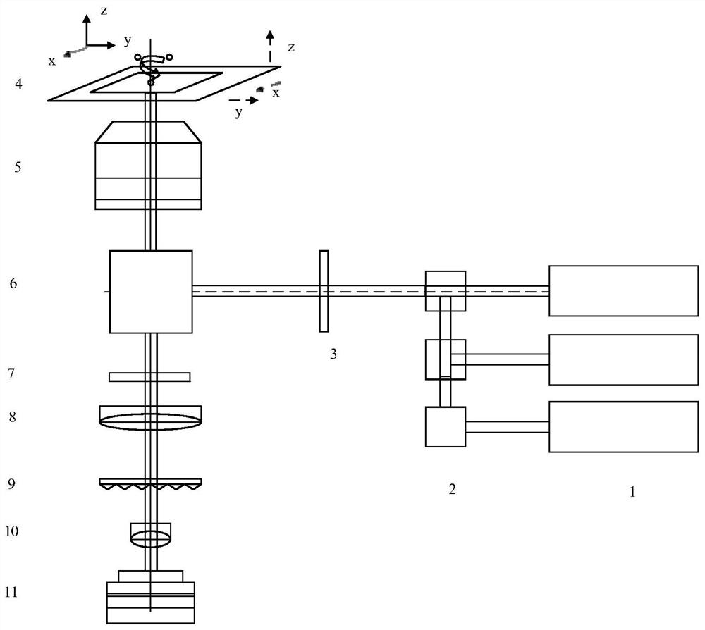
[0044] see first image 3 , image 3 It is a structural block diagram of the multi-color fluorescent microscopic super-resolution imaging system of the present invention. It can be seen from the figure that the present invention is based on light field multi-dimensional information fusion microscopic super-resolution imaging device, including a multicolor fluorescence microscopic imaging system and a single exposure spectral imaging system. The multicolor fluorescent microscopic imaging system includes N sets of different wavelengths Laser 1, N dichroic filters 2, multi-channel narrow-band filters 3, three-dimensional nano-platform 4 for sample placement, microscope objective lens 5, dichroic film 6, mult...


PUM


Abstract

A microscopic super-resolution imaging device and imaging method based on light field multi-dimensional information fusion, the device includes a multi-color fluorescence micro-imaging system and a single-exposure spectral imaging system, and the multi-color fluorescence micro-imaging system includes N sets of different wavelengths The laser, N dichroic filters, multi-channel narrow-band filters, three-dimensional nano-platform for sample placement, microscope objective lens, dichroic film, multi-channel filters and sleeve lens; the single exposure spectrum The imaging system sequentially includes a spatial random phase modulator, a relay amplification imaging system, and a photodetector. The invention can detect and obtain the ultrafine structure of biological living cells with a single exposure, has fast imaging speed, does not require high excitation light power density, and can be applied to real-time super-resolution imaging of living cells. Provide more meaningful raw data for biomedical research.

Description

technical field [0001] The invention relates to fluorescence microscopic super-resolution imaging, in particular to a microscopic super-resolution imaging device and imaging method based on light field multi-dimensional information fusion. Background technique [0002] In optical microscopy imaging, the diffraction-limited resolution is determined by the numerical aperture of the microscopic objective lens and the wavelength of the incident light, and is an objective existence that cannot exceed the theoretical limit resolution. Microscopic super-resolution techniques can be divided into three categories: [0003] 1. Sparse reconstruction technology based on single-molecule localization, representative technologies include light-activated localization microscopy (PALM), stochastic optical reconstruction microscopy (STORM) and fluorescence-activated localization microscopy (FPALM); [0004] 2. Based on point spread function engineering, it is a spatial processing technology ...

Claims


Application Information

Patent Timeline
Patent Type & Authority Patents(China)
IPC IPC(8): G01N21/64G01J3/28G01J3/44G02B21/00
CPCG01N21/6458G01N21/645G01N21/6428G01J3/4406G01J3/2823G02B21/0076G01N2021/6439
Inventor 韩申生童智申刘震涛胡晨昱刘盛盈
Owner SHANGHAI INST OF OPTICS & FINE MECHANICS CHINESE ACAD OF SCI
Features
  • R&D
  • Intellectual Property
  • Life Sciences
  • Materials
  • Tech Scout
  • Unparalleled Data Quality
  • Higher Quality Content
  • 60% Fewer Hallucinations
Learn More