
Imaging and reporting combination in medical imaging

A medical imaging and reporting technology, applied in medical reporting, medical imaging, medical informatics, etc., can solve problems such as inefficient reporting, inconsistency, etc.

Pending Publication Date: 2019-11-01
SIEMENS HEALTHCARE GMBH
View PDF3 Cites 2 Cited by
  • Summary
  • Abstract
  • Description
  • Claims
  • Application Information

AI Technical Summary

Problems solved by technology

This focus does not address inefficiencies in reporting or inconsistencies in reporting

Method used


Image

  • Imaging and reporting combination in medical imaging
  • Imaging and reporting combination in medical imaging
  • Imaging and reporting combination in medical imaging

Examples


Embodiment Construction

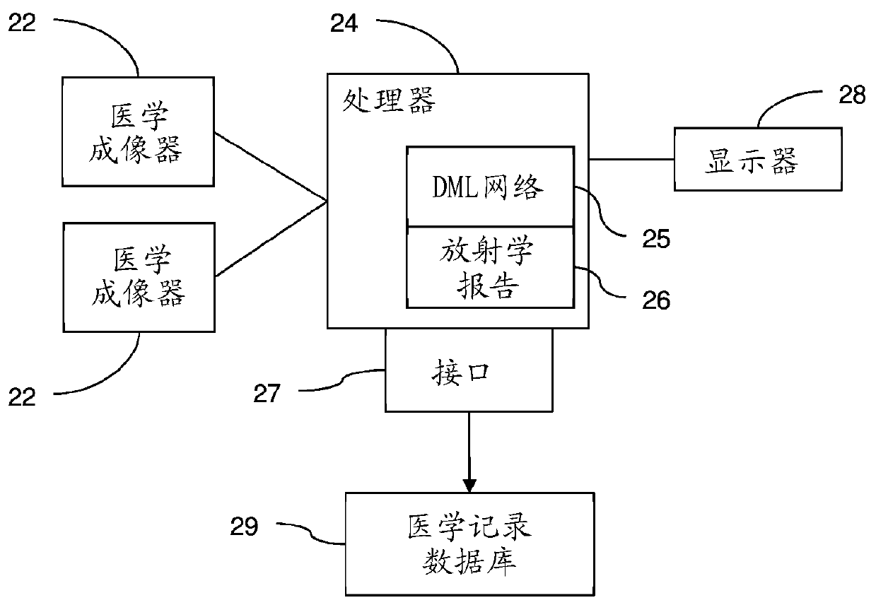
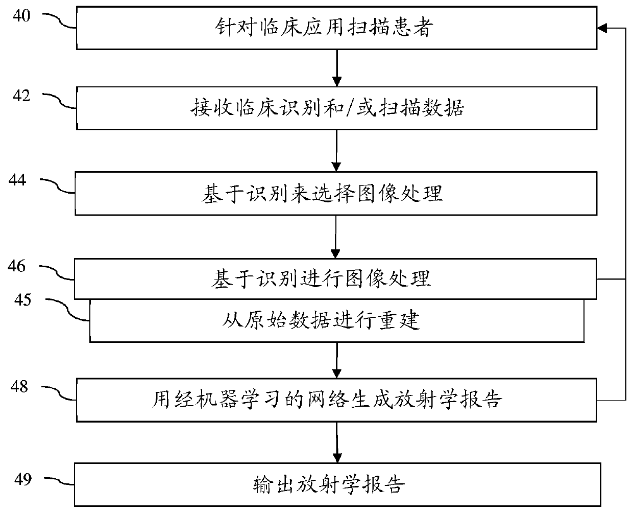
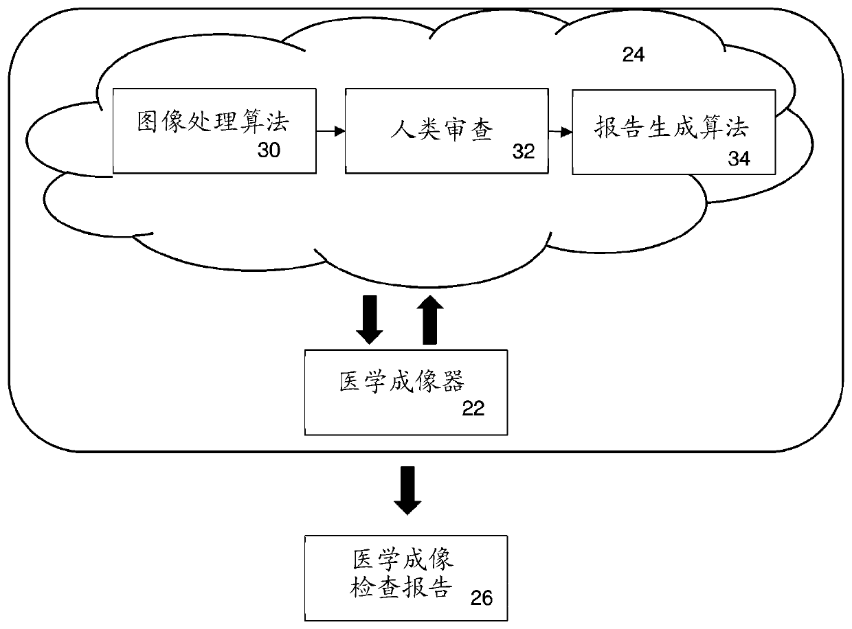
[0014] An integrated medical imaging and reporting device or process scans a subject and produces a clinical report as a final output. Such as figure 1 As shown, the integrated medical imaging and reporting facility includes a medical imaging device 22 and a fulfillment center or processor 24 for interacting to generate a medical imaging exam report 26 . In contrast to a typical medical imaging scanner that produces medical images as its final product, the comprehensive device produces a medical imaging exam report describing the major findings in the patient's imaging exam. The fulfillment center 24 is the medical image reading and reporting infrastructure. Unlike teleradiology operations that are independent of imaging, fulfillment center 24 is specifically configured to operate in a manner that is inherently interconnected with medical imaging equipment 22 . Feedback can be used to control imaging based on ongoing clinical findings from imaging. Feed-forward reconstructi...


PUM


Abstract

The invention relates to imaging and reporting combination in medical imaging. Since the final output for medical imaging is the radiology report, the quality of which is largely dependent on the radiologist, there is a need for a comprehensive system for both medical imaging and reporting. Imaging and radiology reporting are combined. Image acquisition, reading of the images, and reporting are linked, allowing feedback of readings to control acquisition so that the final reporting is more comprehensive. Clinical findings typically associated with reporting may be used automatically to feedback for further or continuing acquisition without requiring a radiologist. A clinical identification may be used to determine what image processing to perform for reading, and / or raw (i.e., non-reconstructed) scan data from the imaging system are provided for integrated image processing with report generation.

Description

technical field [0001] This embodiment relates to medical imaging and reporting from imaging. Background technique [0002] Medical reports, such as radiology reports, are primarily written communications between radiologists, medical professionals, and patients. These reports often contain complex anatomical, technical, and medical information, including images, measurements, and text notation (eg, key findings) providing an analysis or summary of the pathology represented in the images. Its comprehensive interpretation is of great value in the diagnosis, prognosis and treatment of diseases. [0003] Currently, the processes of image acquisition and image reporting are separated. The role of medical imaging scanners is primarily to produce high-quality diagnostic images, while the role of radiologists is to interpret those images and produce radiology reports with key findings. With the focus on value-based care and improving productivity, there is an increased emphasis ...

Claims


Application Information

Patent Timeline
Patent Type & Authority Applications(China)
IPC IPC(8): G16H15/00
CPCG16H15/00G16H30/20G16H30/40G16H50/20G16H50/70G16H40/20G16H10/60G16H80/00
Inventor P.莎马D.科马尼丘
Owner SIEMENS HEALTHCARE GMBH
Learn More