X-ray CT apparatus and computing method for exposure

A technology of exposure dose and calculation method, which is applied in the fields of radiological diagnosis instruments, medical science, diagnosis, etc., can solve the problems of increasing the burden of imaging operators, and achieve the effect of accurate exposure dose value.

Inactive Publication Date: 2004-07-07
GE MEDICAL SYST GLOBAL TECH CO LLC
View PDF1 Cites 7 Cited by
  • Summary
  • Abstract
  • Description
  • Claims
  • Application Information

AI Technical Summary

Problems solved by technology

[0009] On the other hand, the work of measuring the periphery of the imaging area (head circumference or chest circumference) of each patient before imaging and inputting the results to the operation console will place an additional burden on the imaging operator (radiologist, doctor, or nurse, etc.)

Method used


Image

  • X-ray CT apparatus and computing method for exposure
  • X-ray CT apparatus and computing method for exposure
  • X-ray CT apparatus and computing method for exposure

Examples


Embodiment Construction

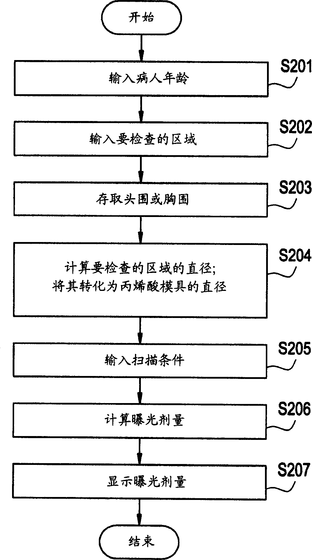
[0019] Several preferred embodiments of the present invention will be described in detail below with reference to the accompanying drawings. Throughout the drawings, the same reference numerals designate the same or similar parts.

[0020]

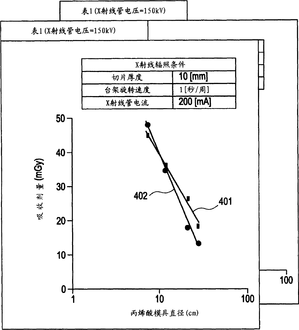
[0021] FIG. 1 shows a system configuration of an X-ray CT apparatus according to one embodiment of the present invention.

[0022] As shown in Figure 1, the X-ray CT equipment includes: a gantry 120, which is used to irradiate the object to be inspected (patient) with X-rays, and detects that the X-ray passes through the object to be inspected; Transmission instruction signal, so that several kinds of settings are activated, and according to the projection (projection) data that is used for displaying from stand 120 output, reconstruct X-ray tomographic image; Carrying device 140, is used for carrying the object of inspection and sending it to the stand Rack 120.

[0023] The platform designated by reference numeral 120 includes a mast...


PUM


Abstract

For the purpose of displaying a more accurate exposure dose value without imposing additional work on an imaging operator, an exposure dose calculating method in an X-ray CT apparatus comprises the steps of: inputting (S201 , 202) the age and a region to be examined of a subject; calculating (S203, S204) the head or chest diameter of the subject based on the age and region to be examined of the subject input at the inputting step, and on statistics on the head or chest circumference of a human body; calculating (S206) an exposure dose of a phantom having a diameter equivalent to the calculated diameter of the head or chest, based on prior information on an exposure dose to a phantom having a predetermined diameter when a predetermined X-ray dose is applied to the phantom; and displaying (S207) the exposure dose value.

Description

technical field [0001] The present invention relates to an X-ray CT (Computed Tomography) apparatus for acquiring a tomographic image of an inspection object with X-ray irradiation. Background technique [0002] X-ray CT equipment generates X-rays around the patient by rotating the X-ray source, and uses X-ray detectors to detect X-rays passing through the patient at different emission angles, and then performs computer processing on the operation console to obtain ( Reconstructing) an image (tomographic X-ray image) of a cross-section (a plane at a slice position, ie, a slice plane) of an inspection object (patient) in an X-ray irradiation area. [0003] When the X-ray CT equipment is used to capture the X-ray tomographic image of the patient, the exposure dose value (CTDI) is displayed in advance on the operation console, which indicates the degree of exposure of the captured image to the patient. [0004] The IEC and FDA recommend using a value measured with a specified ...

Claims


Application Information

Patent Timeline
Patent Type & Authority Applications(China)
IPC IPC(8): A61B6/00A61B6/03
CPCA61B6/542A61B6/583A61B6/03
Inventor 堀内哲也
Owner GE MEDICAL SYST GLOBAL TECH CO LLC
Why Eureka
Try Eureka