Point reconstruction based very large-scale medical image 3D visualization method

A medical imaging and ultra-large-scale technology, which is applied in the field of 3D visualization of ultra-large-scale medical imaging, can solve the problems of not being suitable for real-time visualization of ultra-large-scale data sets, difficulties in 3D real-time processing and display, and many triangles.

CN1632834AInactive Publication Date: 2005-06-29INST OF AUTOMATION CHINESE ACAD OF SCI
0 Cites 19 Cited by

Patent Information

Authority / Receiving Office
CN · China
Patent Type
Applications(China)
Current Assignee / Owner
Publication Date
2005-06-29
Estimated Expiration
Not applicable · inactive patent

Smart Images

  • Figure 1
    Figure 1
  • Figure 2
    Figure 2
  • Figure 3
    Figure 3
Patent Text Reader

Abstract

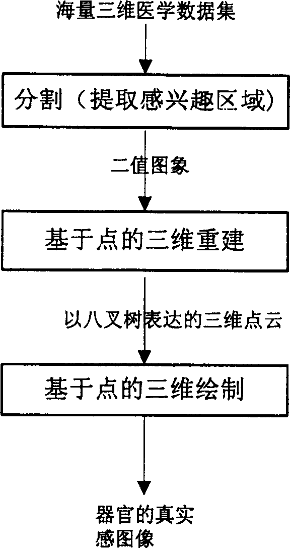
This invention relates to computer graphics and mode identification technique field and in detail to use point recreation for super large scale of medical three-dimensional image visualization and comprises the following steps: first to divide the cared part from the medical two-dimensional slice; second to recreate based on point to extract the cared organic three-dimensional surface and to store it in form of point; third to draw with three-dimension to display the extracted surface mold with the drawing method based on points.
Need to check novelty before this filing date? Find Prior Art

Description

technical field

[0001] The invention relates to the technical fields of computer graphics and pattern recognition, in particular to a method for performing three-dimensional visualization of ultra-large-scale medical images by using point reconstruction. Background technique

[0002] Since the invention of X-rays, modern medical imaging equipment such as CT (Computed Tomography), MRI (Magnetic Resonance Imaging), CR (Computer X-ray Imaging), B-ultrasound, and electronic endoscope have appeared one after another, making traditional medical diagnosis methods appear. a revolutionary change. With the development of modern computer science and technology, medical image processing has emerged as a new interdisciplinary subject, bringing a new dawn to medical diagnosis. However, the traditional way of viewing films is two-dimensional, requiring trained radiologists to view films and make judgments. With the development of computer visualization technology, three-dimensional visual...

Examples

Embodiment

[0070] We apply this method to our self-designed 3D medical image processing and analysis system. The microcomputer-based 3D medical image processing and analysis system 3DMed developed by us is implemented in the computer Windows XP / 2000 / NT / 98 environment, adopts object-oriented design methods and software engineering specifications, and is implemented in C++ language and is oriented to the medical field. 3D image processing and analysis system. This system has rich graphics and image processing and analysis functions, not only has perfect two-dimensional image processing and analysis functions, but also has powerful three-dimensional processing and analysis, network transmission and storage functions. The functions provided by the system include a series of functions such as data input, image data management, 2D processing, 3D data processing, slice reorganization, 3D display, surgical simulation, virtual endoscopy, PACS (image archiving and communication system) and remote ...