Corneal epithelioid cells derived from surface ectodermal cells

a surface ectodermal cell and corneal epithelioid technology, which is applied in the direction of genetically modified cells, peptide sources, peptides, etc., can solve the problems of insufficient donor supply and rejection reaction, low transparency of the sheet using the oral mucosal epithelium, and many limitations in research and use of stem cells, so as to reduce the risk of tumorigenic transformation, short period of time, and favorable

Active Publication Date: 2019-10-29
OSAKA UNIV
View PDF9 Cites 0 Cited by
  • Summary
  • Abstract
  • Description
  • Claims
  • Application Information

AI Technical Summary

Benefits of technology

[0028]No expression of keratin 12 is observed for a conventional cultured oral mucosal epithelial cell sheet, which has been considered to be a cause of its insufficient transparency. According to the present invention, a cell positive for keratin 12 specific to corneal epithelium can be induced from an ectoderm-derived cell, such as an oral mucosal epithelial cell. In addition, the use of the keratin 12-positive cultured oral mucosal epithelial cell as a cell source enables the preparation of a corneal epithelioid cell sheet having properties close to the properties of an corneal epithelial cell compared to those of a conventional cultured oral mucosal epithelial sheet.
[0029]According to the method of the present invention, since a corneal epithelioid cell is directly induced from a somatic cell not through a state of a pluripotent cell, a desired cell can be simply obtained in a short period of time with a reduced risk of tumorigenic transformation compared to that for induction from a pluripotent stem cell. As a result, the method enables a clinically more favorable outcome to be obtained, and is also conducive to the improvement of patient's QOL.

Problems solved by technology

Corneal transplantation through eye donation is performed for intractable corneal epithelial disease; however, it has problems of absolute donor shortage and rejection reaction after transplantation.
However, this method has the following problem: the sheet using the oral mucosal epithelium is low in transparency and weak compared to that using the corneal epithelium since the mucosal epithelium does not differentiate into a complete corneal epithelium.
However, the research and use of the stem cells have many limitations due to ethical problems and there is also a problem of rejection reaction.
Thus, ES cells have not yet been clinically applied.
However, some iPS cells remain as undifferentiated cells even after differentiation induction, and have a risk of tumorigenic transformation after transplanting.
There is also the following problem: the establishment and differentiation induction of iPS cells takes several months or more.

Method used


Image

  • Corneal epithelioid cells derived from surface ectodermal cells
  • Corneal epithelioid cells derived from surface ectodermal cells
  • Corneal epithelioid cells derived from surface ectodermal cells

Examples


example 1

of Corneal Epithelioid Cell from Oral Mucosal Epithelial Cell

[0082]1. Material / Method:

[0083](Cell)

[0084]Immortalized human oral epithelial cells (OKF6 / TERT-1 cells) were used.

[0085](Medium / Culture Condition)

[0086]Cells were cultured under conditions of 5% CO2 and 37° C. in Keratinocyte-SFM to which 25μg / mL bovine pituitary extract (BPE), 0.2 ng / ml epidermal growth factor (EGF), and 0.3 mM CaCl2 were added.

[0087](Vector Construction)

[0088]An all-in-one vector in which PAX6 isoforms, Pa and Pb, their mutants, and PAX6-OCT4-KLF4 were linked using 2A was inserted into pLenti7.3 / V5-DEST vector (Life Technologies). OCT4 and KLF4 were inserted into CSV-CMV-MCS-IRES2-Venus plasmid (RIKEN BIO Resource Center).

[0089]The PAX6 isoforms were hereinafter sometimes referred to as Pa and Pb; KLF4, as K; and OCT4, as O.

[0090](Transduction)

[0091]The pLenti7.3-related vector was introduced into 293FT cells using ViraPower Lentiviral Expression System (Life Technologies) to produce a lentivirus. The CS...

example 2

ion of Genes into Various Cells

[0105]1. Material / Method

[0106](Cell Line)

[0107]Surface ectodermal cell: OKF6 / TERT-1, OKF6 / TERT-2, N / TERT-1, N / TERT-2, HOK

[0108]Neuroectoderm: ARPE-19, SH-SY5Y

[0109]Neural crest: HCEC

[0110]Mesoderm: HUVEC, NHDF-Ad, 293T

[0111]Endoderm: MKN1, HepG2

[0112]iPS cell: 201B7

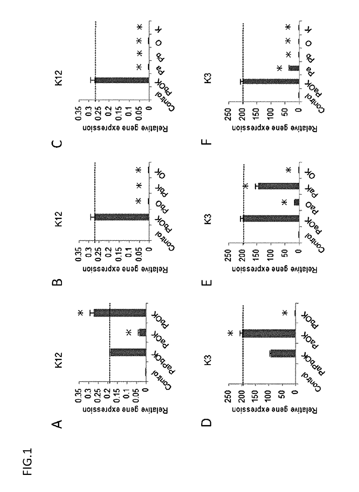
[0113]PAX6 (Pa, Pb), KLF4, and OCT4 were introduced in various combinations into each of the above cell lines as in Example 1, and the expression of K12 and K3 on the resultant cells was confirmed by real-time PCR.

[0114]2. Result

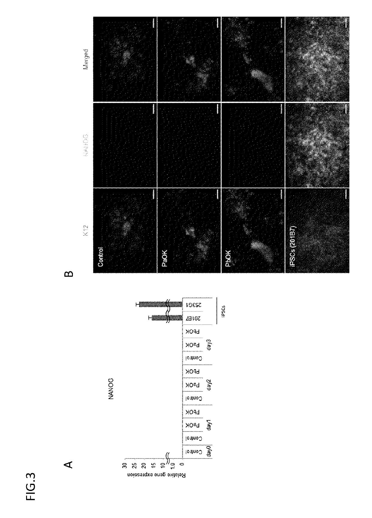
[0115]The results are collectively shown in FIG. 5. K12 / K3 could be efficiently induced mainly on surface ectoderm-derived cells, but were little induced on endoderm- and mesoderm-derived cells and iPS cells. The induction efficiency was high in the surface ectoderm-derived cells having the same developmental origin as corneal epithelium.

example 3

ion of Gene Using All-in-One Vector

[0116]1. Method

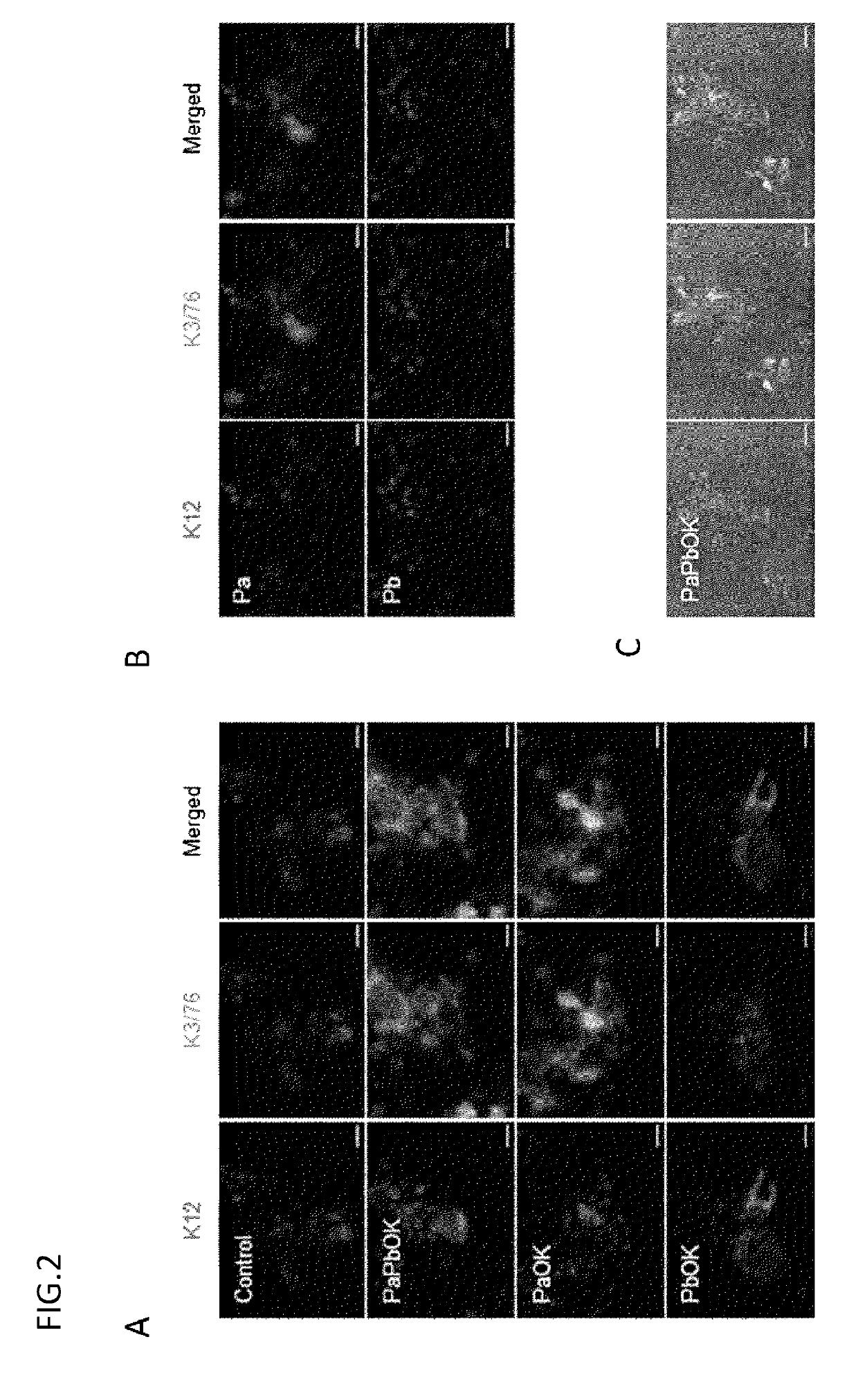
[0117]Using an all-in-one vector, 3 genes (PAX6, OCT4, and KLF4) were liked in the combination of PaOK or PbOK to a lentivirus vector through 2A sequences to prepare each of 2 types of vectors. The vector was transduced into oral mucosal epithelial cells in the same way as in Example 1, and the expression of mRNAs and proteins was evaluated by electrophoresis, real-time PCR, and immunostaining.

[0118]2. Result

[0119]As a result of electrophoresis, the proteins were confirmed to be expressed separated from one another. The results of real-time PCR and immunostaining confirmed that the use of the all-in-one vector provided the sufficient expression of PAX6, OCT4, and KLF4 and thereby enabled the induction of K12 and K3 (FIG. 6).


PUM

PropertyMeasurementUnit
transparencyaaaaaaaaaa
real-time PCRaaaaaaaaaa
structureaaaaaaaaaa

Abstract

The present invention relates to a method for inducing a keratin 12-positive corneal epithelioid cell from a surface ectoderm-derived cell. More specifically, the present invention relates to a method for inducing a keratin 12-positive corneal epithelioid cell, comprising introducing PAX6, KLF4, and OCT4 into a surface ectoderm-derived cell, such as an oral mucosal epithelial cell, and a corneal epithelioid cell induced by the method.

Description

RELATED APPLICATIONS[0001]This application is a National Stage application of PCT / JP2016 / 050619, filed Jan. 12, 2016, which claims priority to Japanese Patent Application No. 2015-004389, filed Jan. 13, 2015, the contents of which are incorporated herein by reference.TECHNICAL FIELD[0002]The present invention relates to a method for inducing a keratin 12-positive corneal epithelioid cell from a surface ectoderm-derived cell. More specifically, the present invention relates to a method for inducing a K12-positive corneal epithelioid cell, comprising introducing PAX6, KLF4, and OCT4 into a surface ectoderm-derived cell, such as an oral mucosal epithelial cell, and a corneal epithelioid cell induced by the method.BACKGROUND ART[0003]Corneal transplantation through eye donation is performed for intractable corneal epithelial disease; however, it has problems of absolute donor shortage and rejection reaction after transplantation. To solve these problems, a therapeutic method using patie...

Claims


Application Information

Patent Timeline
Patent Type & Authority Patents(United States)
IPC IPC(8): C12N5/071C12N15/86C07K14/47
CPCC12N15/86C12N5/0602C07K14/4702C12N2740/15043C12N2506/097C12N2501/603C12N2501/604C12N2501/60C12N5/0621C12N2501/11C12N2510/00C12N5/10
Inventor NISHIDASASAMOTO, YUZURUHAYASHI, RYUHEI
Owner OSAKA UNIV
Why Eureka
Try Eureka