Apparatus and methods for performing brain surgery

a brain surgery and apparatus technology, applied in the field of apparatus and methods for brain surgery, can solve the problems of high invasiveness of brain surgery, difficult diagnosis and treatment of brain conditions, and inadequacies of techniques,

Active Publication Date: 2009-02-19
VYCOR MEDICAL INC
View PDF25 Cites 100 Cited by
  • Summary
  • Abstract
  • Description
  • Claims
  • Application Information

AI Technical Summary

Benefits of technology

[0027]The devices and methods disclosed herein provide numerous advantages in performing brain surgery. Gentle atraumatic dilation of the brain tissue makes it possible to operate further inside the brain than otherwise would be possible utilizing traditional surgical techniques. The disclosed methods and apparatus create an access area to work while simultaneously protecting adjacent brain tissue from inadvertent collateral damage and trauma that might otherwise occur if more traditional surgical techniques were to be utilized. In addition, accessing target tissue through the cannula as contemplated avoids more invasive techniques that involve removing substantial portions of the skull and retracting large portions of the brain to gain access to operate on target tissues. In some cases, the devices and methods may make it possible to operate on target tissue that would, without these devices and methods, otherwise be regarded as inoperable using previously known techniques.

Problems solved by technology

Diagnosis and treatment of conditions affecting the brain are among the most difficult and complex problems that face the medical profession.
Given the difficulty of accessing the brain through the hard bony protective skull the diagnosis and treatment of brain disorders presents unique challenges not encountered elsewhere in the body.
Brain surgery can be highly invasive.
Of course, such techniques are not appropriate for all situations, and not all patients are able to tolerate and recover from such invasive techniques.
It is also known to access certain portions of the brain by forming a hole in the skull, but only limited surgical techniques may be performed through such smaller openings.

Method used


Image

  • Apparatus and methods for performing brain surgery
  • Apparatus and methods for performing brain surgery
  • Apparatus and methods for performing brain surgery

Examples


first embodiment

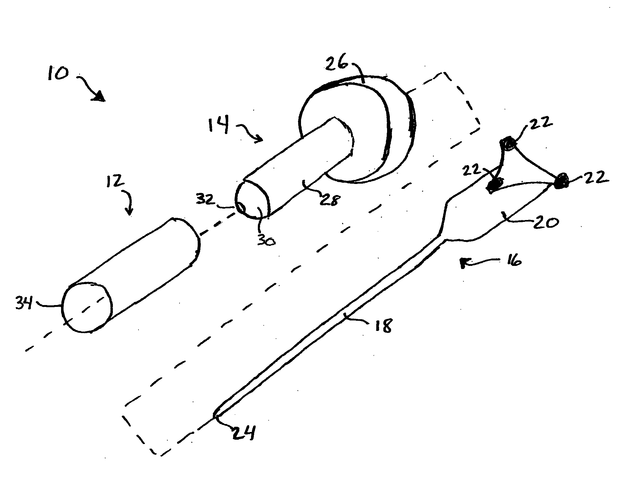
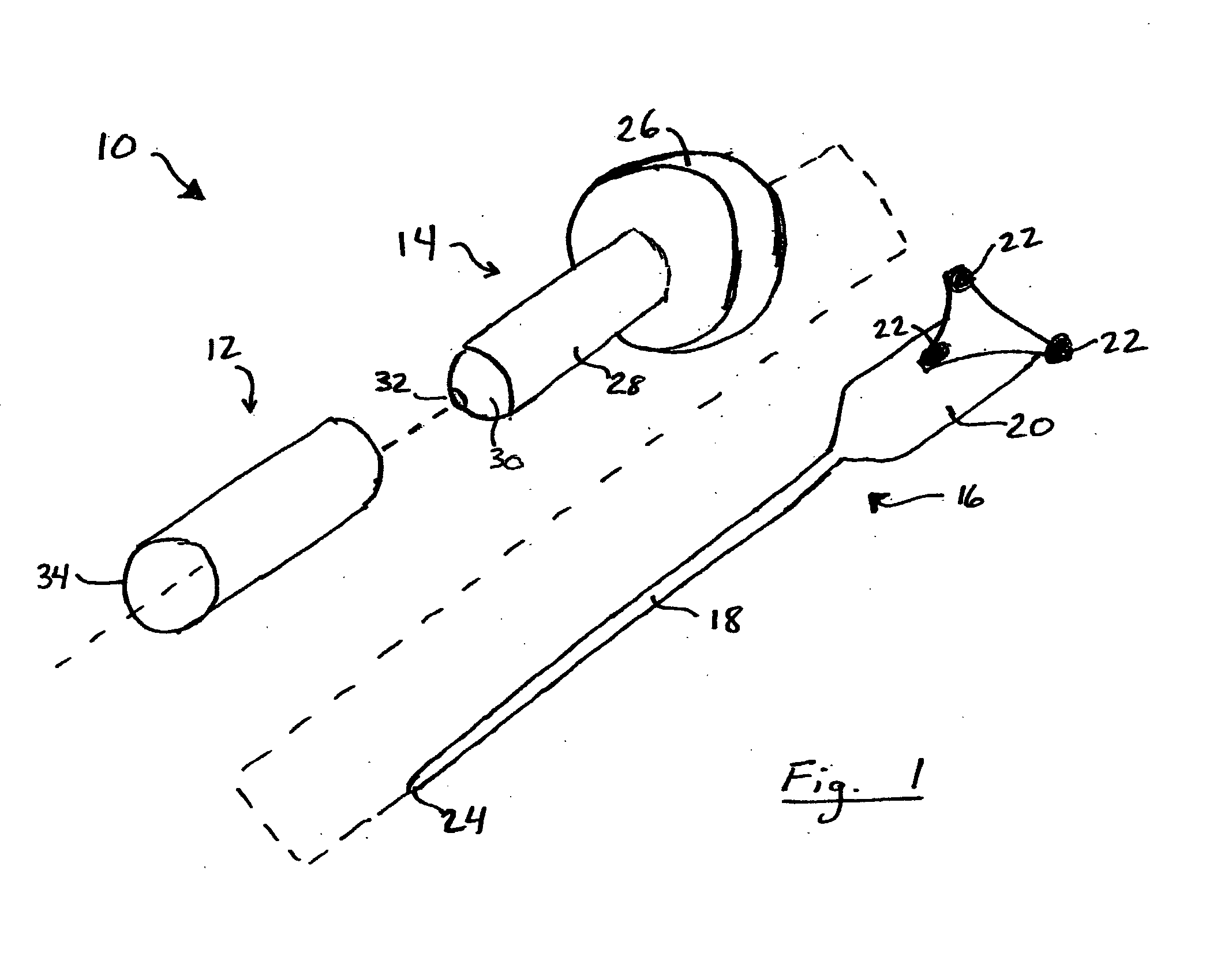
[0052]Referring now to the drawings, FIG. 1 illustrates an apparatus 10 for accessing target tissue within the brain in order to perform brain surgery. The access device includes a cannula 12, a dilating obturator 14 and a stylet or probe 16. Stylet or probe 16 has a small diameter elongated shaft 18, a handle 20 and associated position indicators 22 for a position guidance system. Stylet shaft 18 has a blunt tip 24 that can be inserted into and advanced through brain tissue. In FIG. 1, image guidance position indicators are shown as infrared reflectors of the type use in connection with optical image guidance systems, although other position indicating systems could be used. As shown, the infrared reflectors used with such a system are mounted to the stylet handle in a customary triangular configuration calibrated to identify the tool to the image guidance system. Such imaging systems are available, for example Medtronic Surgical Navigation Technologies (Denver, Colo.), Stryker (Ka...

third embodiment

[0059]FIG. 5 is a perspective view, with parts separated, of an access device 70 for brain surgery. Access device 70 includes cannula 72 with chamfered lead edge 74, and a dilating obturator 76. Dilating obturator 76 includes a handle 78, substantially cylindrical shaft 80 and rounded dilating tip 82, which may be semi-spherical. Access device 70 does not include apparatus for calibrating the position of the dilating obturator with an image guidance system or a stylet or probe for aiding insertion of the dilating obturator.

[0060]FIGS. 6A and 6B illustrate an alternative dilating obturator and cannula assembly which may be used with the foregoing embodiments. More specifically, FIGS. 6A and 6B illustrate an optical dilating obturator which permits visualization as the optical dilating obturator and cannula assembly is inserted into the brain.

[0061]Referring now to FIG. 6A the optical dilating obturator and cannula assembly is configured to include a cannula 82 and a dilating obturato...


PUM


Abstract

Less invasive surgical techniques for performing brain surgery are disclosed in which a dilating obturator and cannula assembly is inserted into brain tissue until the obturator tip and cannula are adjacent to the target tissue. The obturator is removed and surgery is performed through the cannula. In preferred embodiments the obturator and cannula are placed using image guidance techniques and systems to coordinate placement with pre-operative surgical planning. A stylet with associated image guidance may be inserted prior to insertion of the obturator and cannula assembly to guide insertion of the obturator and cannula assembly. Surgery preferably is performed using an endoscope partially inserted into the cannula with an image of the target tissue projected onto a monitor. Dilating obturator structures having a rounded or semi-spherical tip and/or an optical window for visualizing brain tissue during expansion are contemplated.

Description

PRIORITY[0001]This application claims the benefit of the priority of co-pending U.S. provisional application 60 / 622,991, filed Oct. 28, 2004.TECHNICAL FIELD[0002]The present disclosure relates to methods of accessing and performing surgery within the brain.BACKGROUND[0003]Diagnosis and treatment of conditions affecting the brain are among the most difficult and complex problems that face the medical profession. The brain is a delicate soft tissue structure that controls bodily functions through a complex neural network connected to the rest of the body through the spinal cord. The brain and spinal cord are contained within and protected by significant bony structures, e.g., the skull and the spine. Given the difficulty of accessing the brain through the hard bony protective skull the diagnosis and treatment of brain disorders presents unique challenges not encountered elsewhere in the body.[0004]Diagnosis of brain disorders requires clear, accurate imaging of brain tissue through th...

Claims


Application Information

Patent Timeline
Patent Type & Authority Applications(United States)
IPC IPC(8): A61B17/00A61B1/00
CPCA61B17/3431A61B17/3439A61B19/201A61B19/5212A61B2017/00907A61B2017/3456A61B2019/208A61B17/0218A61B19/5244A61B2090/103A61B34/20A61B90/361A61B90/11A61B1/00154A61B5/062A61B17/3423A61B17/3496A61B90/10A61B17/3403A61B2017/00902A61B2017/3425A61B2017/3445
Inventor WILSON, JEFFREY A.
Owner VYCOR MEDICAL INC
Why Eureka
Try Eureka