

Cancer diagnosis based on levels of antibodies against globo h and its fragments
a technology of globo h and antibodies, applied in the field of cancer diagnosis based on levels of antibodies against globo h and its fragments, can solve the problem that the level of these antibodies alone is not a reliable indication of breast cancer
- Summary
- Abstract
- Description
- Claims
- Application Information
AI Technical Summary
Benefits of technology
Problems solved by technology
Method used
Image

Examples
example 1
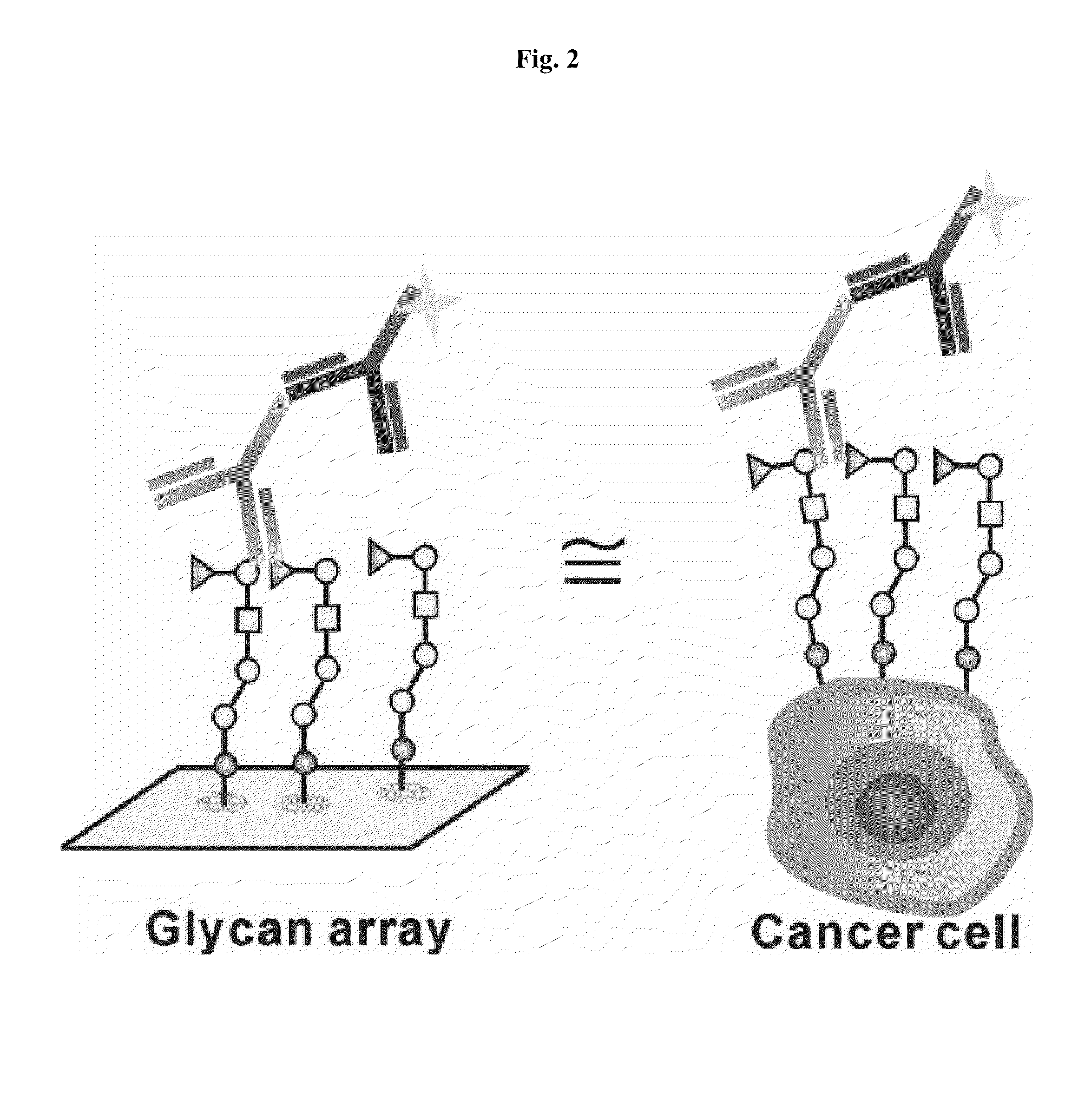
Binding of Antibodies Vk9, Mbr1, and A488 to A Glycan Array
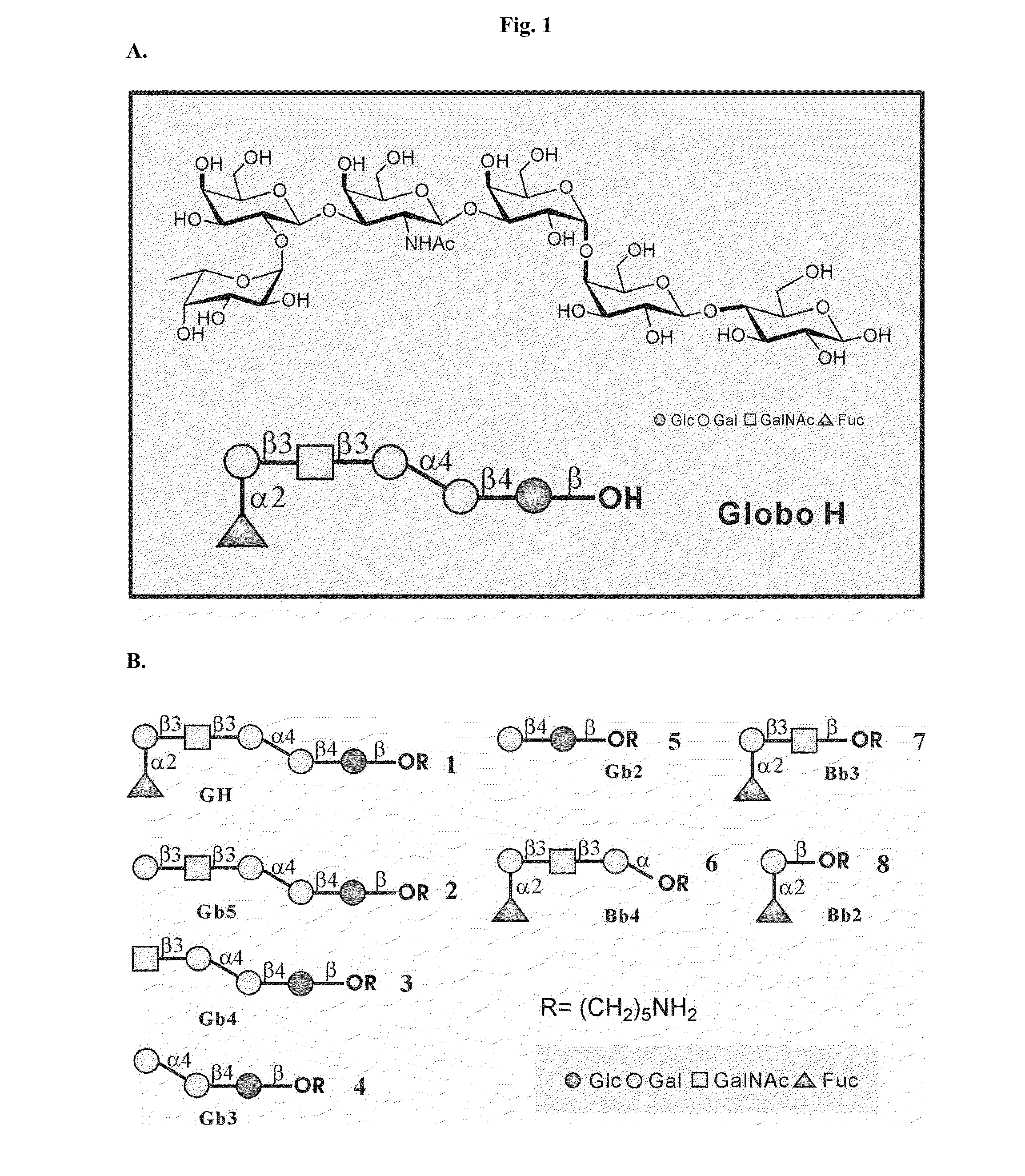
[0015]Oligosaccharides Globo H, Gb5, Gb4, Gb3, Gb2, Bb4, Bb3, Bb2 shown in FIG. 1 were prepared according to the one-pot programmable protocol described in Hung et al., Proc. Natl. Acad. Sci. USA 103-15-20 (2006). These oligosaccharides were covalently attached onto a NHS-coated glass slide, which was purchased from Nexterion H slide (SCHOTT North America), by the standard microarray robotic printing technology described in Hung et al. and Blixt et al., Proc. Natl. Acad. Sci. USA 101:17033-17038 (2004). More specifically, an aliquot from a stock solution (80 μM) of each oligosaccharide was placed on the glass slide in 16-row format, two rows for each oligosaccharide.
[0016]The following three antibodies were used in this study:[0017]Mbr1, a mouse IgM anti-Globo H monoclonal antibody,[0018]VK-9, a mouse IgG anti-Globo H monoclonal antibody, and[0019]A488, anti-mouse / human Gb5 monoclonal antibody.
[0020]Each of the antibodies wa...
example 2
Use of A Glycan Array for Detecting Antibodies Against Globo H and Its Fragments in Breast Cancer Patients
[0022]Plasma samples from breast cancer patients and healthy individuals were diluted 1:20 with 0.05% Tween 20 / 3% BSA / PBS buffer (pH 7.4) and incubated with the glycan array slide described in Example 1 above in a humidifying chamber with shaking for 1 h. After being washed with 0.05% Tween 20 / PBS buffer (pH 7.4), PBS buffer (pH 7.4), and water, each for three times, the slide was incubated with Cy3-conjugated goat anti-human IgM or IgG antibody in the humidifying chamber with shaking for 1 h. The slide was then washed three times with 0.05% Tween20 / PBS buffer (pH 7.4), three times with PBS buffer (pH 7.4), and three times with H2O. After being dried, the slide was scanned at 595 nm (for Cy3-conjugated secondary antibody) with a microarray fluorescence chip reader (ArrayWorx microarray reader).
[0023]As shown in Tables 1 and 2 below, the level ratios of Globo H-bound IgG / Gb5-boun...
example 3
Use of Glycan Array for Monitoring Immune Responses Induced by Globo H Vaccine
[0024]Mice (6-week-old female BALB / c mice, BioLASCO, Taiwan) were immunized subcutaneously with the Globo H-KLH vaccine (Optimer Pharmaceuticals, Inc., San Diego, Calif.) once every week for three weeks. Control mice were injected with phosphate buffer saline (PBS). Serum samples were collected from the treated mice 10 days after the last immunization. These samples were subjected to serial dilution at 30, 120, 240, 480, 960, and 1920 folds and the titers of anti-Globo H antibodies were examined in the diluted serum samples using the glycan array slide described in Example 1 above (3.5×10⁻¹⁴ mol of oligosaccharides per spot) or by conventional ELISA (coated with 1.28×10⁻¹⁰ mol of Globo H per well).
[0025]The Globo H-KLH vaccine induced secretion of anti-Globo H antibodies in the immunized mice. See Table 3 below. It has also been found that the glycan array assay described above is much more sensitive as co...
PUM
| Property | Measurement | Unit |
|---|---|---|
| pH | aaaaa | aaaaa |
| structures | aaaaa | aaaaa |
| structure | aaaaa | aaaaa |
Abstract
Description
Claims
Application Information