Optical instrument and technique for cancer diagnosis using in-vivo fluorescence emission of test tissue

a cancer diagnosis and fluorescence emission technology, applied in the field of detection of cancer, can solve the problems of still being difficult to identify cancerous tissue by direct in-vivo examination, and requiring sophisticated and expensive instruments capable of detection

Inactive Publication Date: 2001-07-03
WOLFE WILLIAM L
View PDF13 Cites 53 Cited by
  • Summary
  • Abstract
  • Description
  • Claims
  • Application Information

AI Technical Summary

Benefits of technology

is that such diagnostic instrument be simple and inexpensive, so that it can be used by primary care physicians.

Problems solved by technology

In spite of the progress illustrated by the prior art, it still remains difficult to identify cancerous tissue by direct in-vivo examination.
The use of monochromatic or narrow bandwidth for excitation and emission light (such as the 20 nm bandwidth taught by Alfano et al.) requires sophisticated and expensive instrumentation capable of detecting very low light intensities and of functioning with low signal-to-noise ratios.

Method used


Image

  • Optical instrument and technique for cancer diagnosis using in-vivo fluorescence emission of test tissue
  • Optical instrument and technique for cancer diagnosis using in-vivo fluorescence emission of test tissue
  • Optical instrument and technique for cancer diagnosis using in-vivo fluorescence emission of test tissue

Examples


Embodiment Construction

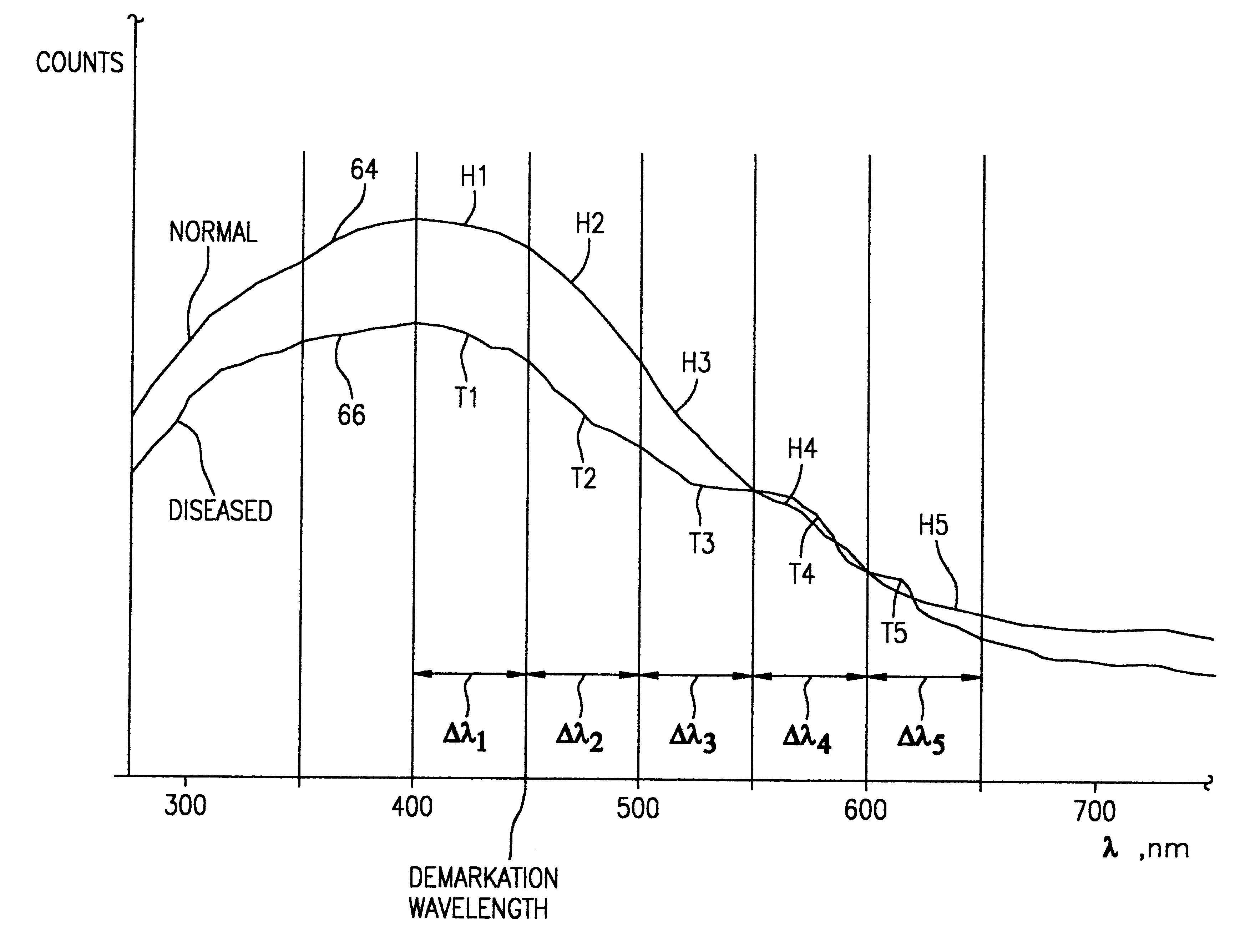
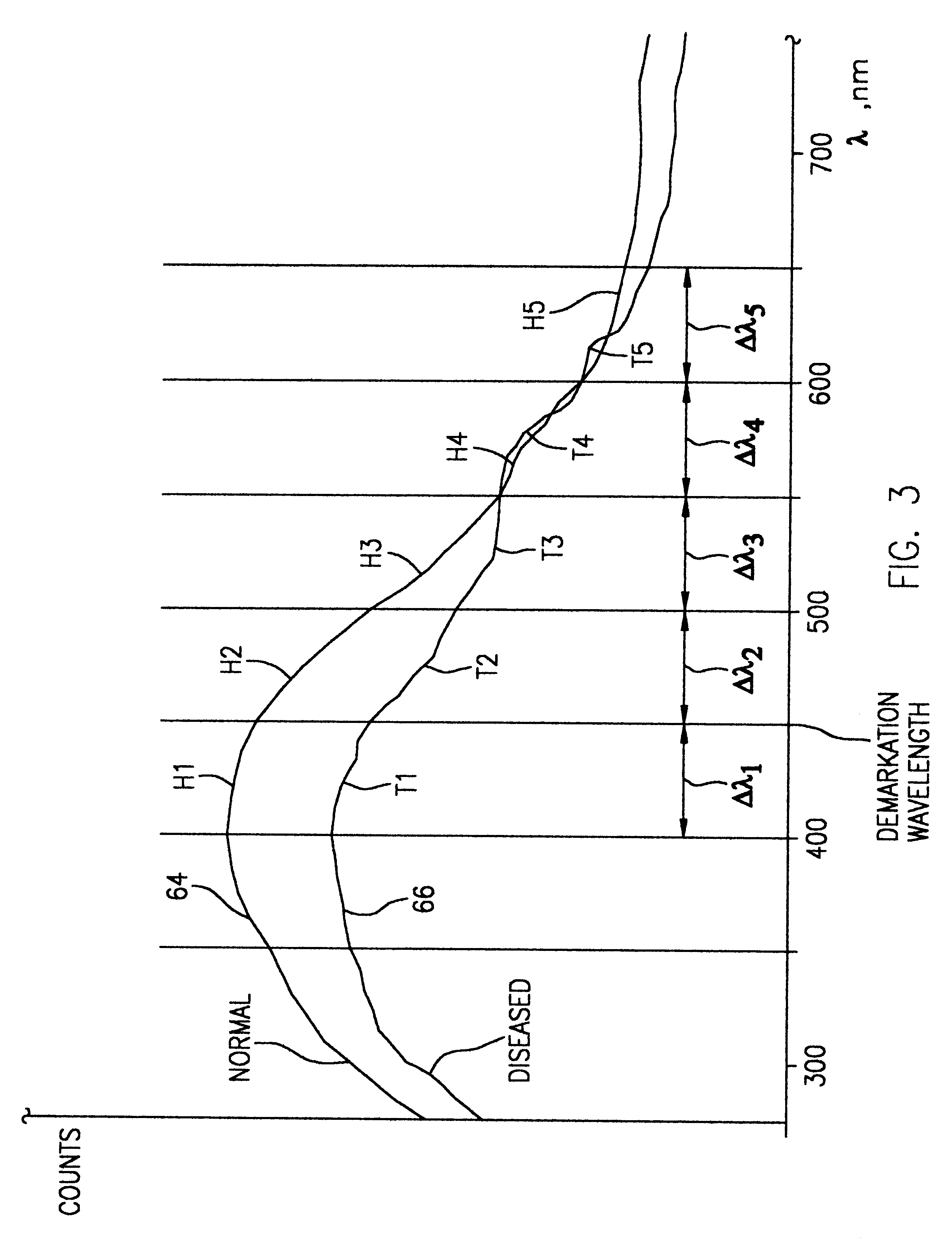
The heart of this invention lies in the recognition that by monitoring wider fluorescent-emission bandwidths than previously done more information is collected at each intensity measurement regarding differences between the emission spectra of cancerous and benign cells. In addition, stronger signals are generated that make it possible to implement the diagnostic technique of the invention with simpler and less expensive technology that can be incorporated in a self-contained, manually-operated device. Finally, this invention discloses a more versatile and discriminating approach for distinguishing the fluorescent emissions of cancerous cells from that of benign tumors and healthy tissue.

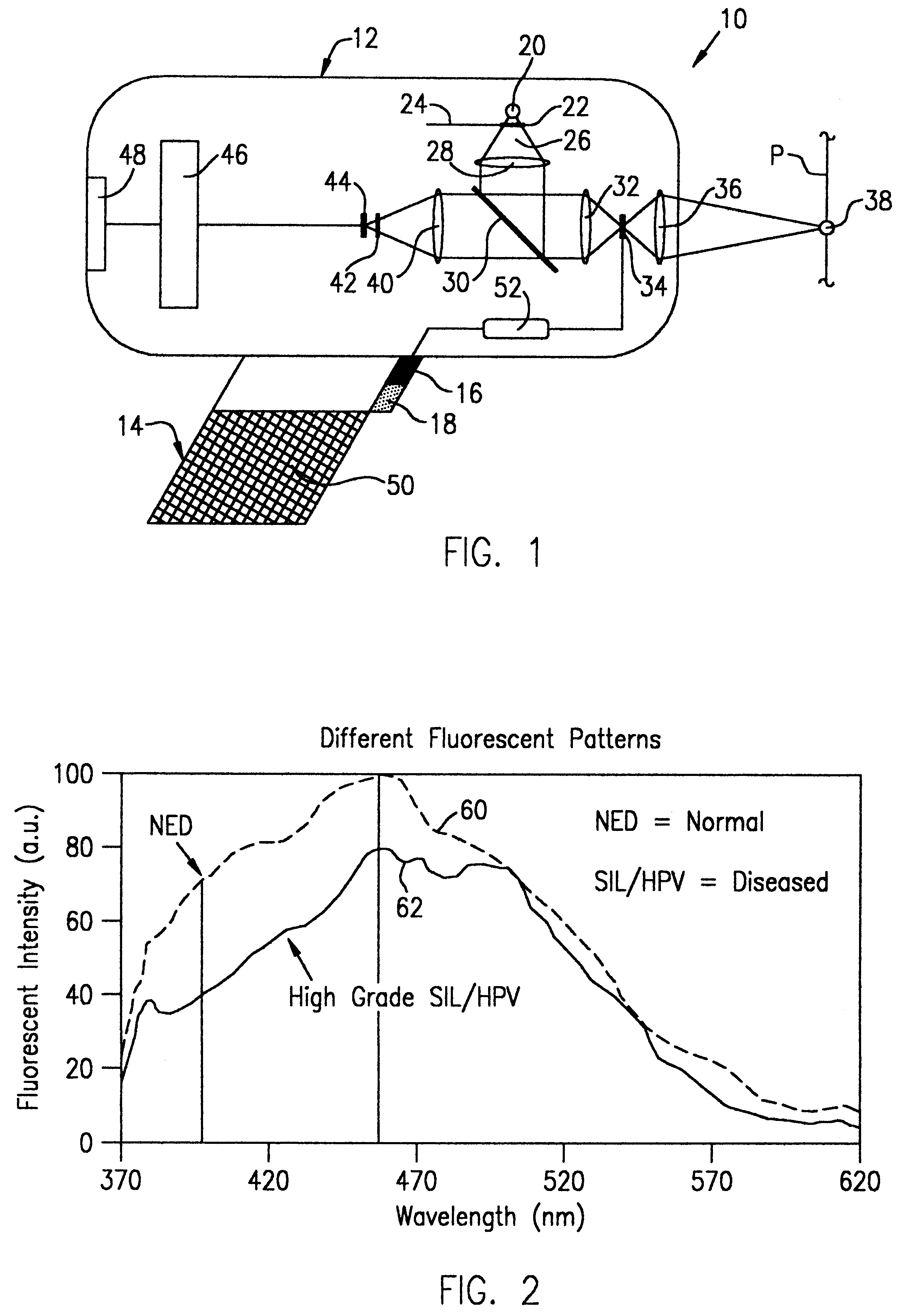
Referring to the drawings, wherein like reference numerals and symbols are used to identify like parts, FIG. 1 illustrates in schematic form a detection instrument 10 according to one embodiment of the invention. The drawing shows the functional components of the instrument 10 as they would appear i...


PUM


Abstract

A hand-held, small, lightweight instrument is disclosed that contains a light source capable of producing radiant energy in the spectral range between approximately 370 and approximately 410 nm, and an optical direction system for irradiating a target tissue by producing an illuminated spot thereon. A collection system is provided for receiving and directing fluorescent emissions in the frequency range between 450 and 700 nm returned from the target area to a detector. A processing system is used to determine the state of the tissue by using at least two and up to five spectral bands, preferably each being larger than 45 nm. Pairs of ratios of fluorescent intensities are compared to identify cancerous cells. An alphanumeric, false-color image and/or audio signal is immediately provided to inform a user of the state of the tissue.

Description

1. Field of the InventionThis invention pertains in general to the detection of cancer in various types of human tissue and, more particularly, to a novel procedure and instrumentation for in-vivo detection of cancerous cells using fluorescence excitation in tissue that is optically accessible for examination.2. Description of the Related ArtSeveral techniques and instruments have been described by investigators for the purpose of detecting cancerous cells. For example, U.S. Pat. No. 5,062,431 to Potter (1991) utilizes in-vivo fluorescence detection of abnormal tissue by irradiating a photosensitized diagnostic region simultaneously with at least two excitation wavelengths. Fluorescent light emitted from the diagnostic region is detected and a signal is generated relating to the intensity of the fluorescence. A difference signal between the two wavelengths is used to distinguish the fluorescence of normal tissue from that of diseased cells.In U.S. Pat. No. 5,106,387 (1992), Kittrell...

Claims


Application Information

Patent Timeline
Patent Type & Authority Patents(United States)
IPC IPC(8): A61B5/00
CPCA61B5/0059
Inventor WOLFE, WILLIAM L.
Owner WOLFE WILLIAM L
Why Eureka
Try Eureka