
Technique and device for laminar osteotomy and laminoplasty
- Summary
- Abstract
- Description
- Claims
- Application Information
AI Technical Summary
Benefits of technology
Problems solved by technology
Method used
Image

Examples
Embodiment Construction
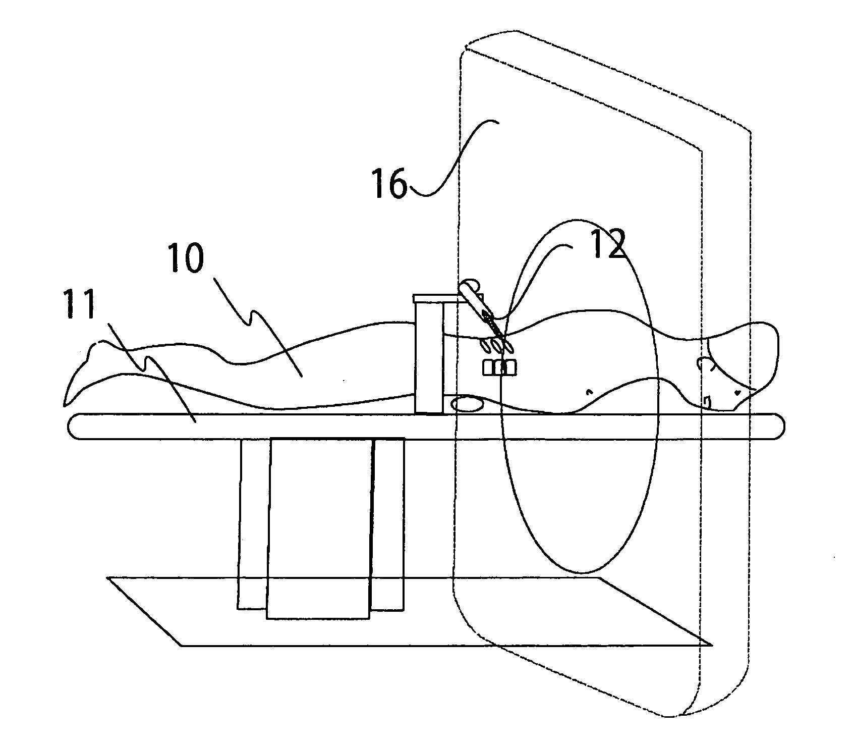
[0041]Preferred embodiments, methods and aspects of surgical implantable devices to be used in dividing of the spinal lamina and elevation of the posterior wall of the spinal central canal are described in this section. This reshaping of the lamina, or laminoplasty results in the creation of more space for the neuronal elements in the spinal central canal. This method is of particular benefit since it can be performed through a small incision under CT, MRI, or other image guidance. Furthermore, the technique can be applied to the cervical, thoracic, and lumbar levels of the spine of any vertebrate.
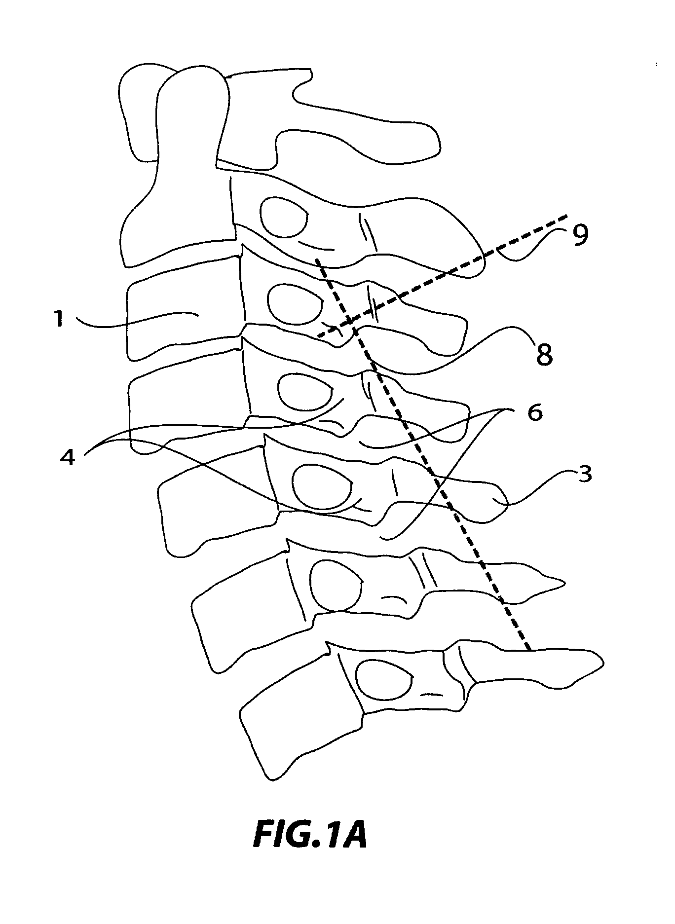
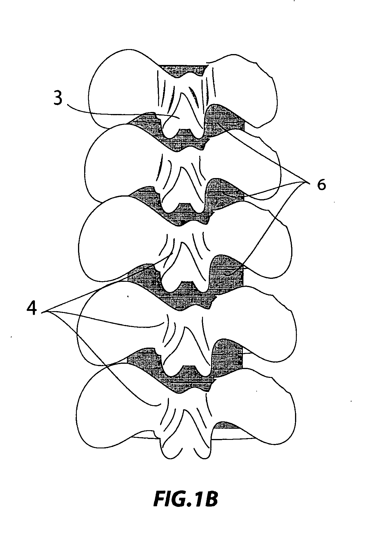
[0042]FIGS. 1-2 show lateral, posterior, and axial views of cervical and lumbar vertebrae respectively. Thoracic vertebra are not drawn as they are similar in the anatomical region relevant to this description. The vertebral bodies 1 create the anterior wall of the central canal 2. The spinous process 3 is a midline bony prominence where the lamina 4 meet. This process elevates the divided...
PUM
 Abstract
Description
Claims
Application Information