Nerve stimulation biopsy device

a biopsy device and nerve stimulation technology, applied in the field of nerve stimulation biopsy devices, can solve the problems of affecting the safety of the biopsy procedure, and affecting the patient's health, so as to achieve the effect of safe biopsy

Inactive Publication Date: 2016-09-15
KLIOT MICHEL +7
View PDF3 Cites 3 Cited by
  • Summary
  • Abstract
  • Description
  • Claims
  • Application Information

AI Technical Summary

Benefits of technology

[0010]The present disclosure provides an advantageous biopsy device that decreases the likelihood of damage to functioning nerve fibers by assisting the user in distinguishing between nerve fibers and tumorous tissue. More particularly, this disclosure provides a nerve stimulation biopsy device whose tip is configured to deliver a precise electrical stimulus (stimulus amplitude, frequency and duration could be controlled) to help localize the cannula in relation to nerve tissue and prevent damage to functioning nerve fibers.
[0011]The biopsy device in accordance with the present invention has the following advantages. First, the nerve stimulation biopsy device has a tip that is capable of delivering a precise electrical stimulus to distinguish between functioning and tumorous nerves, and to prevent damage to functioning nerve fibers. Second, the stimulation signal emitted is capable of being adjusted and controlled in accordance with patient responses, allowing the user to determine the exact proximity to healthy nerve tissue.
[0013]In another aspect, this disclosure provides a method for safely performing a biopsy procedure on a biopsy site of a patient. The method includes inserting a distal end of the cannula assembly into the biopsy site, and introducing via the cannula assembly a stimulus signal of a predetermined strength into the biopsy site. The method further includes receiving feedback from the patient and removing the distal end of the cannula assembly from the biopsy site if said feedback is associated with a positive physical response or continuing with the procedure if said feedback is associated with a negative physical response, wherein said positive physical response is characterized by pain, sensation or muscle reflex and said negative physical response is characterized by an absence of pain, sensation and muscle reflex, and removing the target tissue from the biopsy site

Problems solved by technology

Pathology frequently involves peripheral nerves, often embedded in deep and difficult to access locations.
Currently, biopsies of nerve and associated pathology, such as nerve tumors (e.g., Neurofibromas and Schwannomas), are performed using a biopsy device that cannot identify or distinguish functioning nerve fibers from pathological nerve or tumor tissue.
As a result, biopsies often damage functioning nerve tissue, resulting in long lasting pain and even loss and / or altered sensory and / or motor function.
This presents additional risks to the patient and is often unnecessary as these tumors are rarely malignant.
While there is no device in the market that addresses the specific need in any way, any improvement will compete with the current state of the art technology.
For example, while percutaneous image guided biopsy devices using ultrasound, CT, or MRI, assist in guiding the path for incision, they cannot distinguish between functioning nerve fibers and pathological tissues.
Current guided methods have disadvantages.
An MRI guided biopsy is an expensive and lengthy procedure.
An ultrasound guided biopsy has a number of deficiencies, such as attenuation of the sound beam, and obstruction of visualization.
CT guided biopsies expose patients and surgeons to radiation and may be especially harmful for women and children.
However, because the vibration assisted biopsy device cannot distinguish between functioning nerve fibers and tumorous tissue, the nerve fibers are still at risk.

Method used


Image

  • Nerve stimulation biopsy device
  • Nerve stimulation biopsy device
  • Nerve stimulation biopsy device

Examples


Embodiment Construction

[0020]For the following defined terms, these definitions shall be applied, unless a different definition is given in the claims or elsewhere in this disclosure. As used in this disclosure and the appended claims, the singular forms “a”, “an”, and “the” include plural referents unless the content clearly dictates otherwise. As used in this disclosure and the appended claims, the term “or” is generally employed in its sense including “and / or” unless the content clearly dictates otherwise.

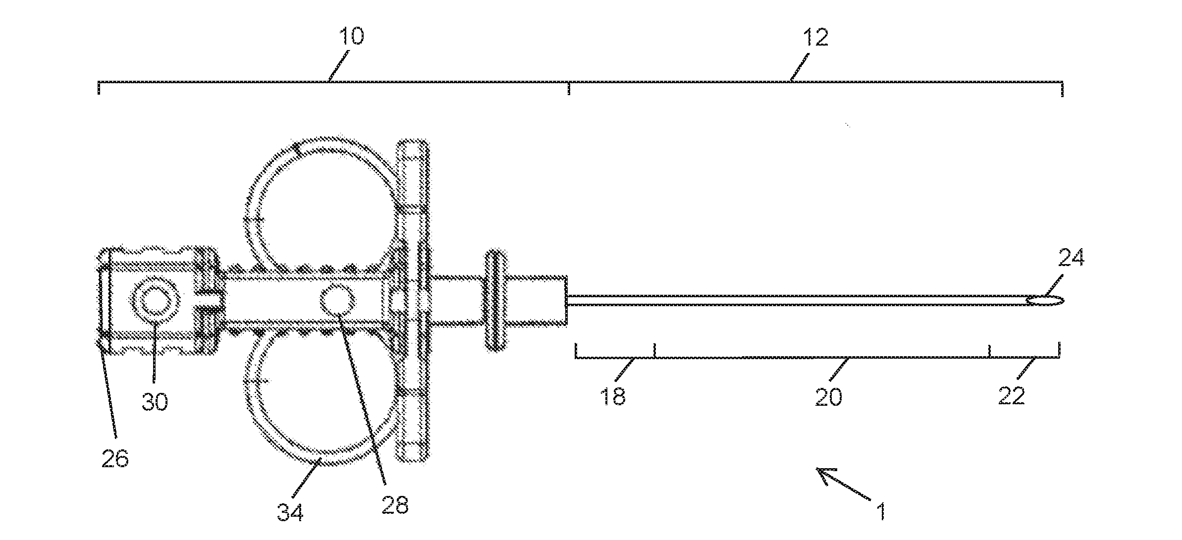
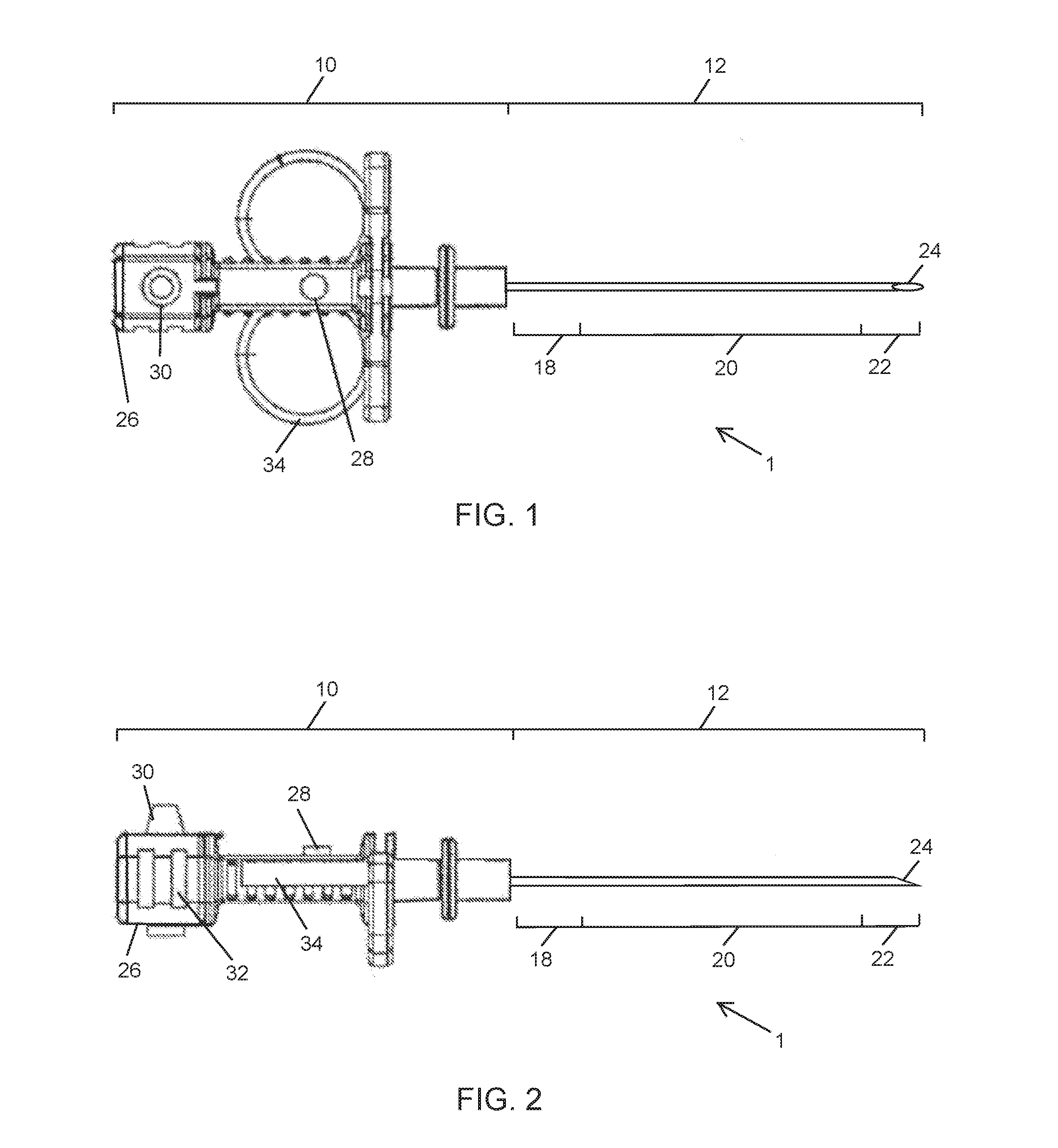
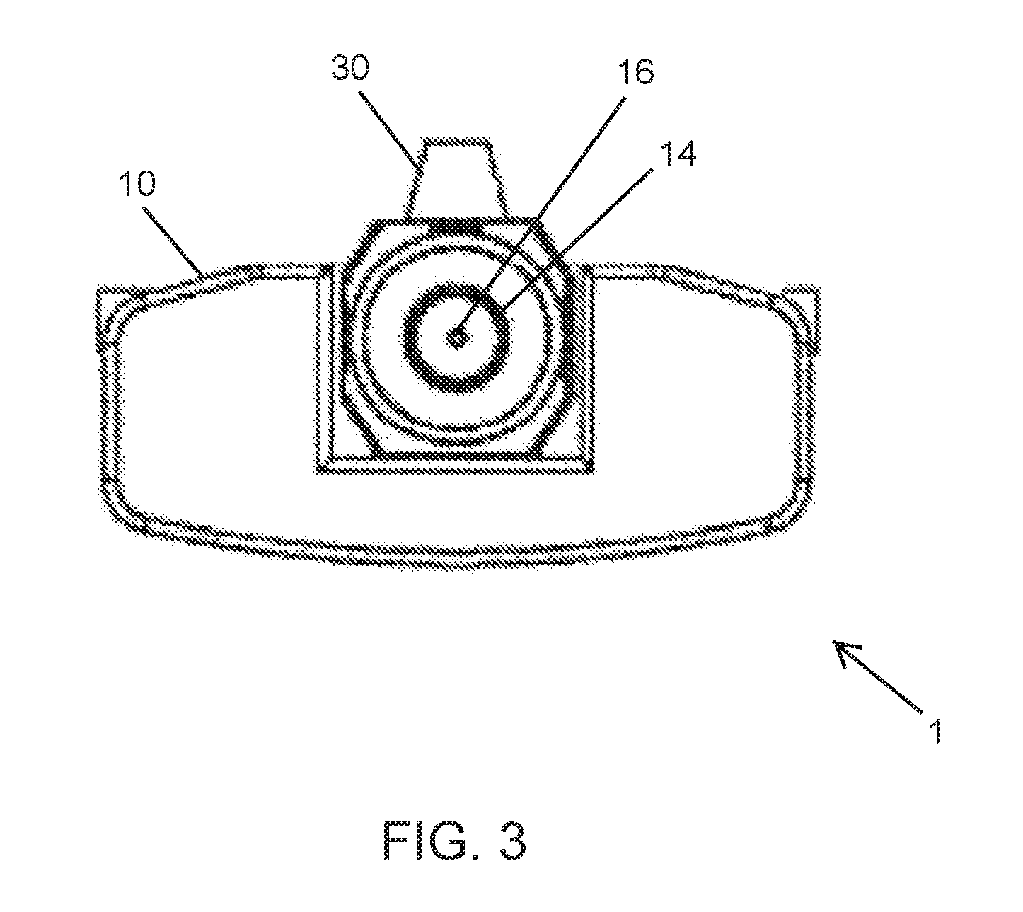
[0021]While the present invention will be described in the specific context of a peripheral nerve biopsy device, the invention is not limited to just peripheral nerve biopsy instruments. The present invention may be used in conjunction with a variety of medical or other instruments and is effective for use in not only peripheral nerve biopsy retrieval procedures, but also with other instruments to perform other tissue removal procedures, or other procedures and associated instruments as would be appre...


PUM


Abstract

A nerve stimulation biopsy device configured to deliver a precise and adjustable stimulus signal into a biopsy site. The stimulus signal is used to distinguish between functioning nerve fibers and target tissue. Also disclosed is a method for safely performing a biopsy procedure on a biopsy site of a patient, which utilizes introducing a stimulus signal to the biopsy site.

Description

CROSS-REFERENCE TO RELATED APPLICATION[0001]This application claims the benefit of U.S. Provisional Patent Application Ser. No. 62 / 130,989, filed Mar. 10, 2015, the disclosure of which is hereby incorporated by reference in its entirety.BACKGROUND OF THE INVENTION[0002]1. Field of the Invention[0003]The present subject matter relates generally to a nerve stimulation biopsy device. More specifically, the present invention relates to a biopsy device possessing a tip configured to deliver an electrical stimulus to distinguish between functioning nerve fibers and target tissue, such as tumorous tissue.[0004]2. Discussion of the Prior Art[0005]Surgery is being performed more rapidly and safely, through smaller incisions, using a variety of technologies, including intraoperative imaging and endoscopic instrumentation. Pathology frequently involves peripheral nerves, often embedded in deep and difficult to access locations.[0006]Currently, biopsies of nerve and associated pathology, such a...

Claims


Application Information

Patent Timeline
Patent Type & Authority Applications(United States)
IPC IPC(8): A61B10/02A61B5/04
CPCA61B5/04001A61B10/0233A61B5/1104A61B5/4029A61B5/4824A61B5/4893
Inventor KLIOT, MICHELBODE, JOHNHEIFERMAN, MICHAELKHANNA, RYANMA, YUNLIPREM, KOMALMESSANA, NICKRUSSI, COLIN
Owner KLIOT MICHEL
Why Eureka
Try Eureka