
Apparatus and method for x-ray treatment
a radiation therapy apparatus and x-ray technology, applied in the direction of diagnostics, therapy, instruments, etc., can solve the problems of difficult to achieve rapidity and high resolution together, apparatus cannot pursue the moving treatment object part in real time, and it is difficult to improve the rapidity any further in the radiation therapy apparatus. to achieve the effect of accurate irradiation to the lesion
- Summary
- Abstract
- Description
- Claims
- Application Information
AI Technical Summary
Benefits of technology
Problems solved by technology
Method used
Image

Examples
Embodiment Construction
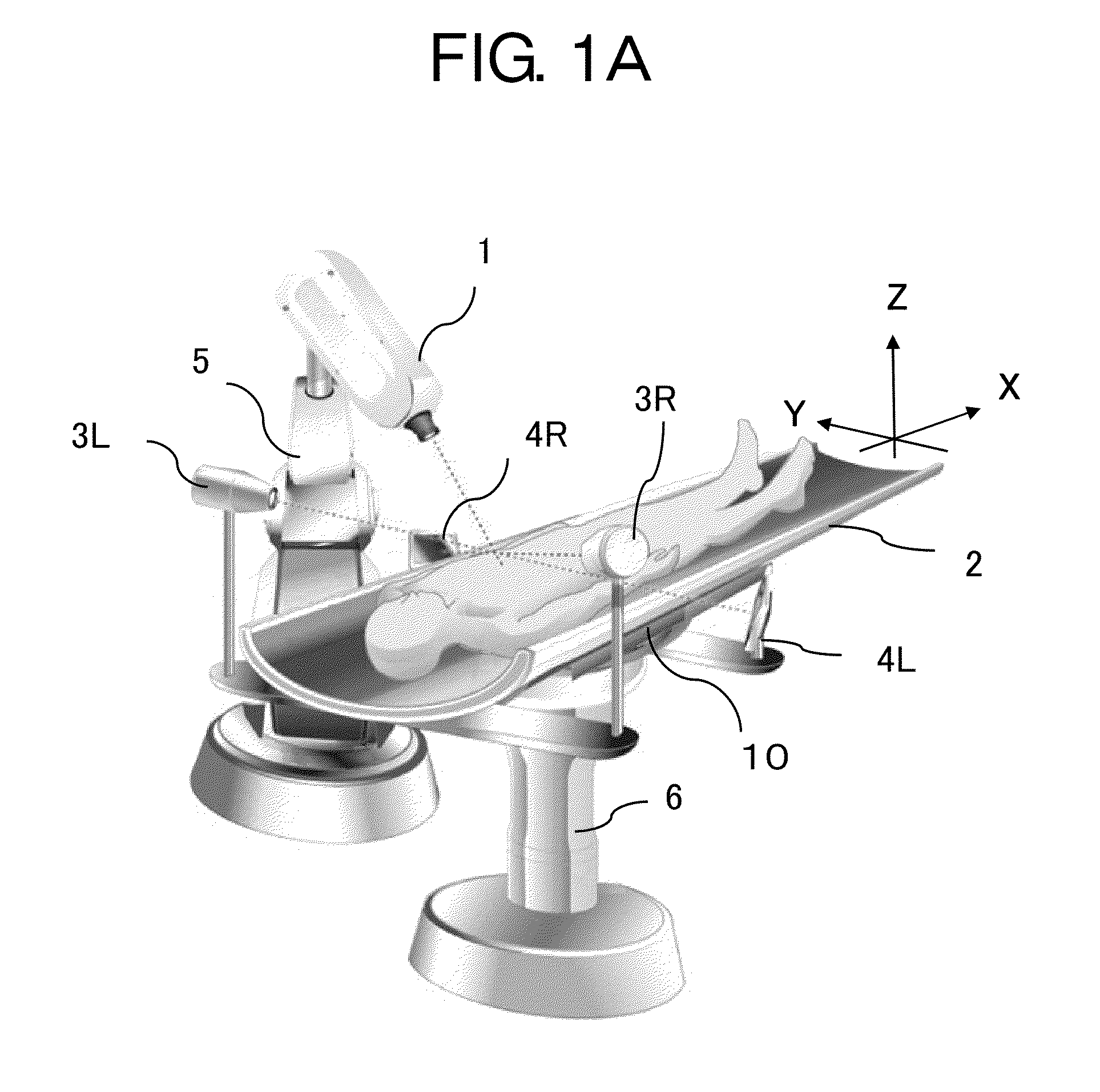
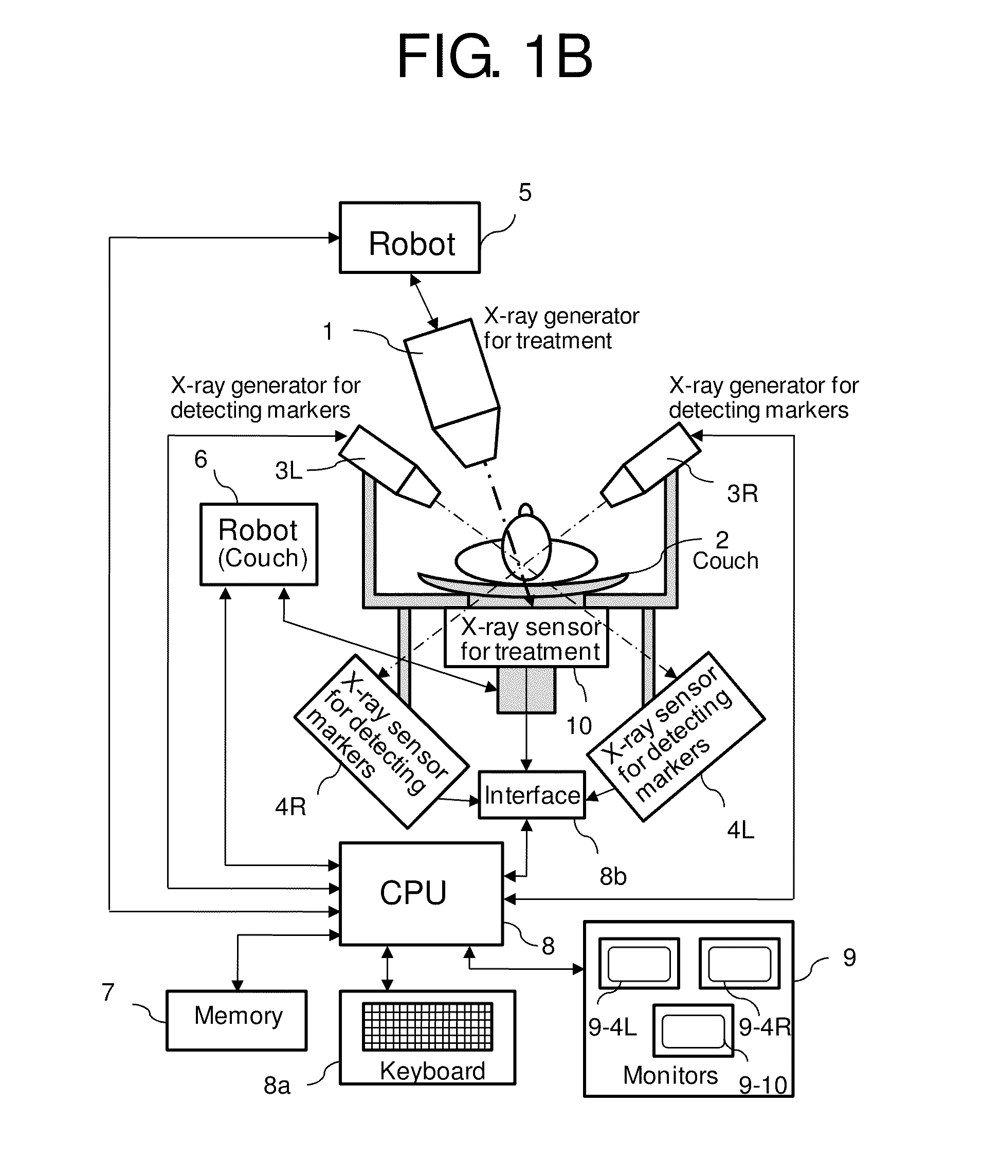
[0033]FIG. 1A shows a perspective view showing the X-ray treatment apparatus in accordance with an embodiment of the present invention, and FIG. 1B shows a block diagram of the X-ray treatment apparatus. The following detailed description is described about the case that the lesion is a tumor.
[0034]The marker is fixed in the vicinity of a tumor in the patient while keeping the predetermined position to the couch. The material of the marker is heavy metal such as Au or Pt which is safe for the human body and absorbs X-rays well.
[0035]The marker sensor includes two low energy X-ray generators 3L, 3R which are installed in the couch 2 and are turned to the tumor to detect the position of a marker, and two image sensors 4L, 4R corresponding to each low energy X-ray generator. The marker sensor collects the information about the tumor which keeps the predetermined position to the marker and the three-dimensional position of the marker to the couch 2.
[0036]The position of couch 2 is chang...
PUM
 Abstract
Description
Claims
Application Information