
Method for treatment of a vascular lesion

a vascular lesion and treatment method technology, applied in the field of vascular lesions treatment, can solve the problems of blood vessel, inability to repeat treatments as many times as required, and long-term effects, and achieve the effect of accurately identifying the contour of vascular lesions

Active Publication Date: 2022-11-15
BARD PERIPHERAL VASCULAR
View PDF32 Cites 0 Cited by
  • Summary
  • Abstract
  • Description
  • Claims
  • Application Information

AI Technical Summary

Benefits of technology

The present invention is a better way to figure out the shape of a blood vessel blockage using ultrasound imaging. The method involves using a special marker in the blood to help identify the type of blockage. This information is then used to tune the treatment for the blockage with a non-invasive device called HIFU. The treatment involves using a high-frequency ultrasound wave to destroy or dissolve the blockage. This method helps to accurately identify and treat blood vessel blockages.

Problems solved by technology

However, these treatments are invasive and / or ionizing, creating long-term effects.
Moreover, these treatments may not be repeated as many times as required to fully destroy the targeted cells.
The difficulty in applying HIFU to lesion or plaque removal in a blood vessel is that the content of the lesion or plaque is not known, e.g., the lesion may be fatty, calcified, or fibrous, or a combination thereof.

Method used


Image

  • Method for treatment of a vascular lesion
  • Method for treatment of a vascular lesion
  • Method for treatment of a vascular lesion

Examples


Embodiment Construction

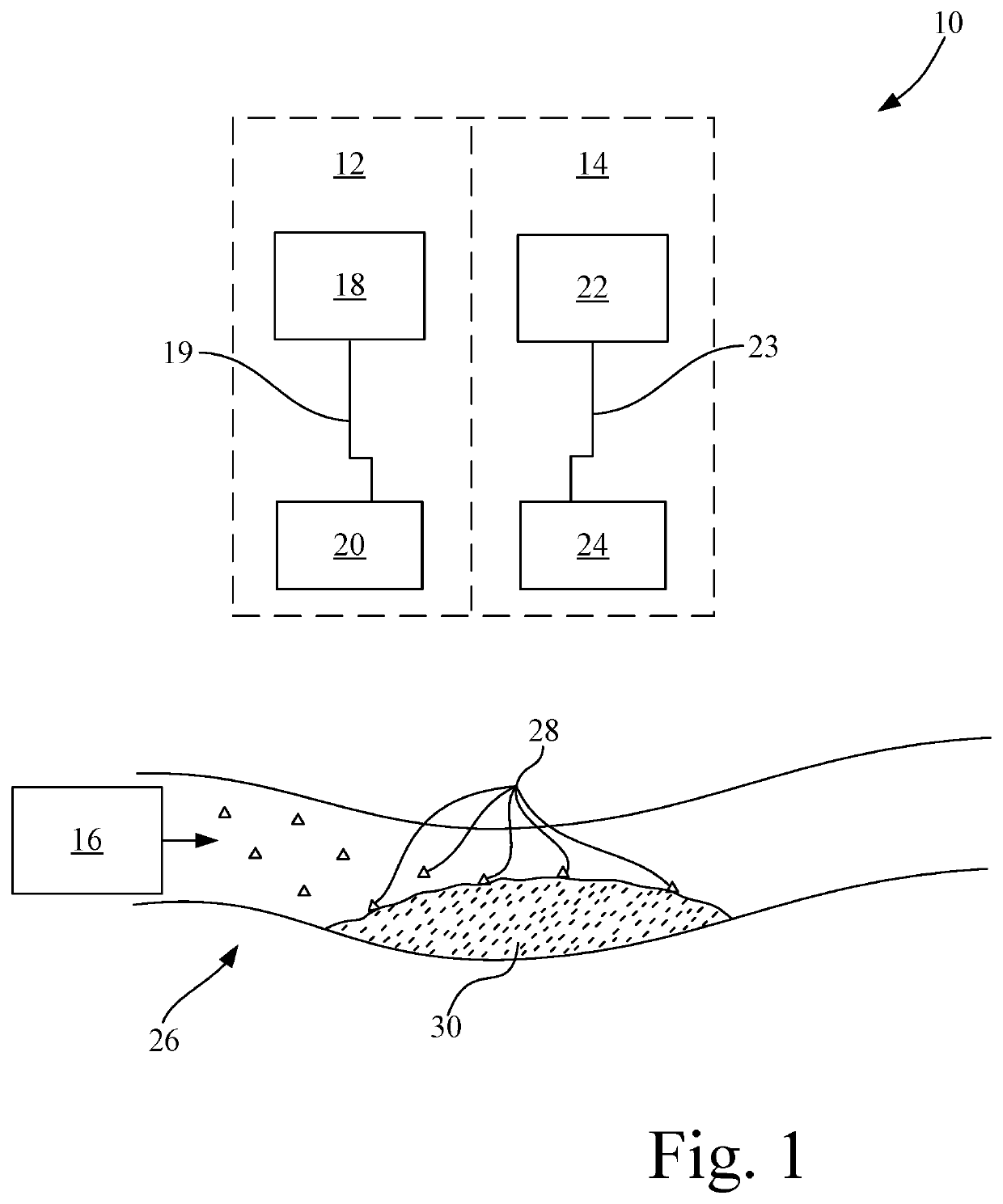
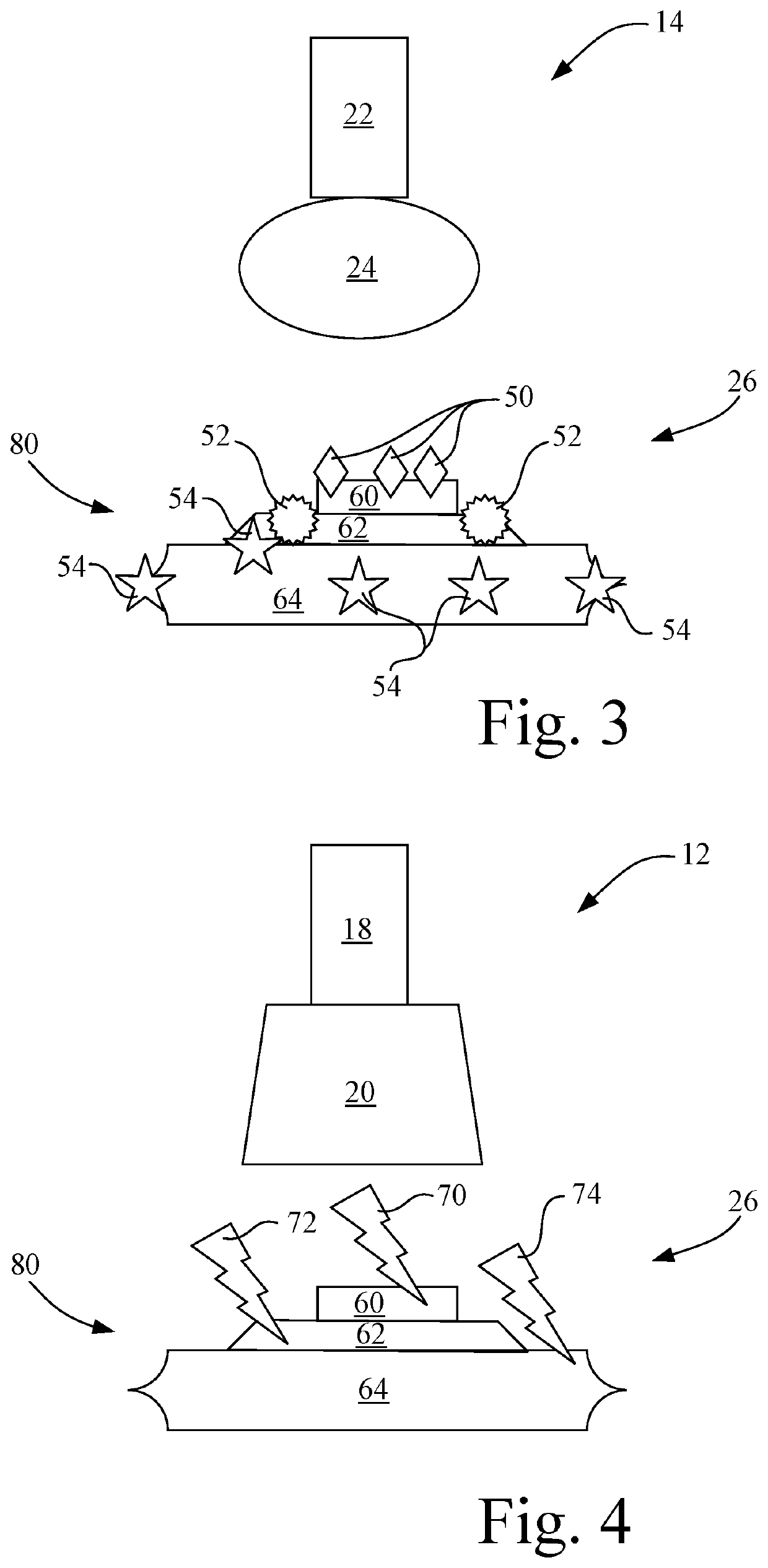
[0016]Referring now to the drawings, and more particularly to FIG. 1, there is shown a system 10 for use in treating a vascular lesion in accordance with an aspect of the invention. System 10 includes a non-invasive high intensity focused ultrasound (HIFU) system 12, a non-invasive ultrasound imaging system 14, and a biomarker insertion device 16.

[0017]Non-invasive HIFU system 12 includes a HIFU generator 18 coupled to a HIFU device 20, e.g., by a first multi-conductor cable 19. In the present embodiment, HIFU device 20 is an HIFU probe. HIFU generator 18 is operable to generate a HIFU emission. HIFU device 20 receives the HIFU emission from HIFU generator 18. The frequency of the HIFU emission may be tuned at the HIFU device 20, e.g., by tuning an oscillator / crystal circuit in HIFU device 20. HIFU device 20 applies the HIFU emission with the tuned frequency to a vascular lesion 30 in a vascular region 26 to break up the vascular lesion.

[0018]HIFU device 20 is operable to tune the f...


PUM


Abstract

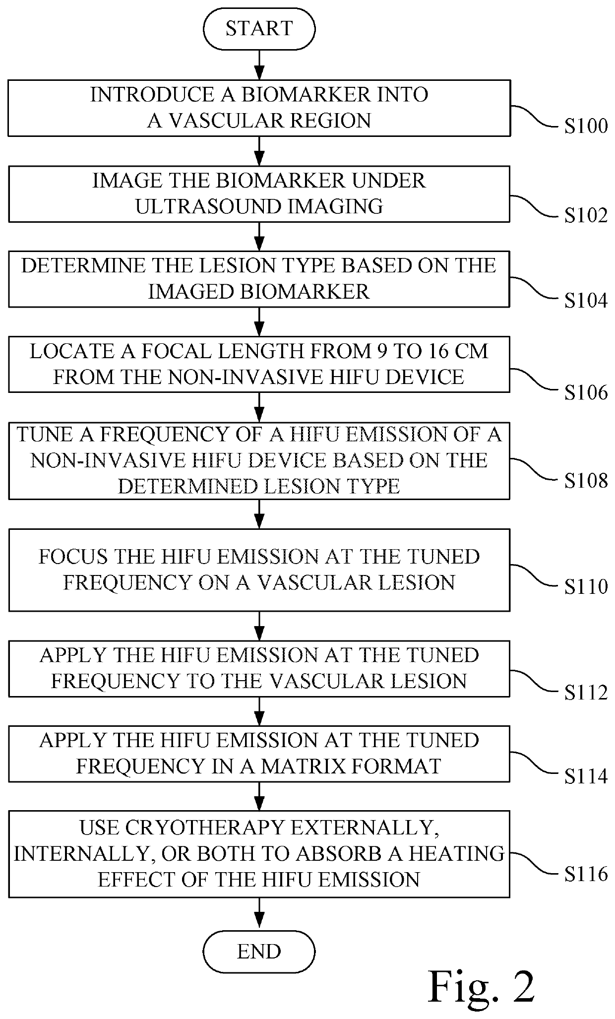
A method for treatment of a vascular lesion. A biomarker insertion device introduces a biomarker into a vascular region. The biomarker binds to a lesion type of a plurality of lesion types. Under ultrasound imaging, an image brightness of an imaged biomarker differs based on the lesion type of the plurality of lesion types to which the biomarker binds. An ultrasound imaging probe of an ultrasound imaging system images the biomarker and produces an image brightness of the imaged biomarker. The lesion type is determined based on the image brightness of the imaged biomarker. A frequency of a high intensity focused ultrasound emission of a non-invasive high intensity focused ultrasound device is tuned based on the determined lesion type. The tuned frequency is applied to the vascular lesion, resulting in the destruction of the vascular lesion.

Description

CROSS-REFERENCE TO RELATED APPLICATIONS[0001]None.TECHNICAL FIELD[0002]The present invention relates generally to treatment of vascular lesions, and, more particularly, to a method of treatment of vascular lesions based at least in part on the lesion type.BACKGROUND ART[0003]Laser ablation, microwave coagulation, and radiofrequency ablation may be used in treating benign and malignant tumors, hemostasis, uterine fibroids, and other conditions. However, these treatments are invasive and / or ionizing, creating long-term effects. Moreover, these treatments may not be repeated as many times as required to fully destroy the targeted cells.[0004]It is known to use High Intensity Focused Ultrasound (HIFU) lithotripsy in kidney stone applications, cellulite treatment, and tumor therapy. The difficulty in applying HIFU to lesion or plaque removal in a blood vessel is that the content of the lesion or plaque is not known, e.g., the lesion may be fatty, calcified, or fibrous, or a combination t...

Claims


Application Information

Patent Timeline
Patent Type & Authority Patents(United States)
IPC IPC(8): A61B8/08G06T7/00
CPCA61B8/085G06T7/0012G06T2207/10132G06T2207/30096G06T2207/30101
Inventor SAMPATH KUMARAN, RANJANI
Owner BARD PERIPHERAL VASCULAR
Learn More