Imaging device for dental instruments and methods for intra-oral viewing

Inactive Publication Date: 2012-02-16
DENTSPLY SIRONA INC
View PDF27 Cites 212 Cited by
  • Summary
  • Abstract
  • Description
  • Claims
  • Application Information

AI Technical Summary

Benefits of technology

[0007]It has been discovered that dental professionals can benefit from adding image viewing capability to several standard dental instruments such as a drill, a curette, a scaler, an extractor, a surgical instrument, a mirror and a toothbrush. A viewing device is provided where all camera components such as illumination, imag

Problems solved by technology

The use of handheld dental instruments such as drills and scalers are impeded by the fact that the area to be treated can be hard to reach and that the instrument itself can block the optimal field of view of the area to be treated.
Although mirrors are routinely used by dental professionals to circumvent this situation, they are both a nuisance by using up the free hand of the dental professional and hard to interpre

Method used


Image

  • Imaging device for dental instruments and methods for intra-oral viewing
  • Imaging device for dental instruments and methods for intra-oral viewing
  • Imaging device for dental instruments and methods for intra-oral viewing

Examples


Embodiment Construction

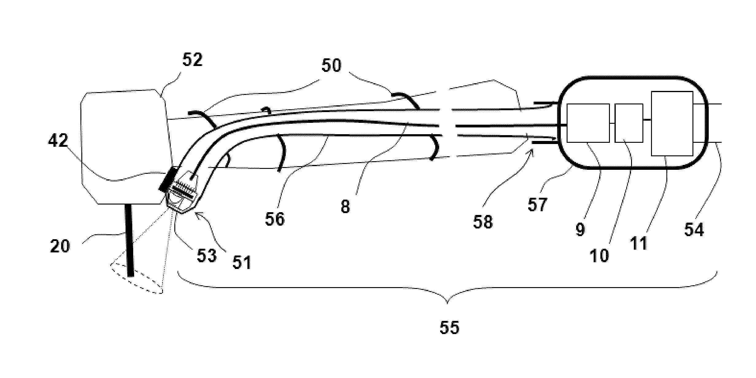
[0036]Referring to the drawings and for illustrative purposes, the present invention can be embodied by the device shown in FIG. 1 which illustrates the attachments 50 of the imaging device 55 to a dental instrument such as a drill 52 with a working tip such as a bur 20. Attachment of the imaging device 55 to the dental instrument 52 can be achieved by a mounting device 42 composed of an attachment body casing made of materials such as silicon or plastic and comprising a means to easily attach, position and detach the imaging device on a dental instrument. The electrical wires 8 allowing transmission between the optical device head 51 and control unit 57 can be secured to the dental instrument 52 with an easy clipping mechanism 50 whereas the optical device head 51 can be secured to the dental instrument head 52 by a permanent magnet found on the mounting device 42. Magnetic mounting of the device head to the instrument head is advantageous as it allows for optimal positioning of th...


PUM


Abstract

The present application relates to an apparatus and method in the field of dentistry and in particular to optical tools for diagnosis and imaging. It is a problem of the prior art that the use of fibre optic components yield a bulky dental camera with poor resolution. It is an object of the present invention to provide an imaging device (55) for dental instruments comprising a chamber adapted to be secured to the head of a dental instrument and for receiving and removing an imaging device (1), the chamber also adapted for isolating said imaging device from said dental instrument without compromising sterility of the dental instrument, comprising an optical window (53). The imaging device is easily removable from the dental instrument and electronic image detection occurs at the instrument head.

Description

FIELD OF THE INVENTION[0001]The present invention relates to an apparatus and method in the field of dentistry and in particular to optical tools for diagnosis and imaging.BACKGROUND OF THE INVENTION[0002]The use of handheld dental instruments such as drills and scalers are impeded by the fact that the area to be treated can be hard to reach and that the instrument itself can block the optimal field of view of the area to be treated. Although mirrors are routinely used by dental professionals to circumvent this situation, they are both a nuisance by using up the free hand of the dental professional and hard to interpret due to image reversal. Nevertheless, it is still common for dental professionals to use handheld mirrors to visualize certain areas of a patient's mouth. It is known in the art to provide dental professionals with a camera for intra-oral use to eliminate the need for handheld mirrors.[0003]Oral cavity cameras are used in the field of dentistry to allow dental profess...

Claims


Application Information

Patent Timeline
IPC IPC(8): A61B1/24
CPCA61B1/00087A61B1/00096A61B1/00101A61B1/05A61B1/051A61B1/053H04N5/3577A61C1/088A61L2/202A61L2/206A61L2202/181A61L2202/24H04N5/2256A61B1/247H04N23/56H04N25/617
Inventor KARAZIVAN, NAIMERTL, THOMASGUARAGNO, KENNETH R.NOVAK, EUGENE J.
Owner DENTSPLY SIRONA INC
Why Eureka
Try Eureka