Ventriculoperitoneal shunt with distal balloon

a technology of ventriculoperitoneal shunt and balloon, which is applied in the direction of balloon catheter, wound drain, etc., can solve the problems of increased icp, serious neurological problems, and 40% failure rate of vp shunt within two years

Inactive Publication Date: 2016-10-13
UNIV OF IOWA RES FOUND
View PDF4 Cites 0 Cited by
  • Summary
  • Abstract
  • Description
  • Claims
  • Application Information

AI Technical Summary

Problems solved by technology

Such CSF buildup typically occurs in the ventricles inside the brain, which cause the ventricles to enlarge and compress the brain against the skull, leading to increased ICP.
This increase in ICP can cause serious neurological problems, including death.
According to medical statistics, VP shunts are the most common neurological procedure performed in the United States, yet these VP shunts have a 40% failure rate within two years, primarily due to catheter migration and / or infection.
For example, the distal catheter extending into the abdominal cavity can accidentally back out from the peritoneum, which causes the CSF to collect under the skin without reabsorption.
Catheter migration or pull out is particularly a problem in children due to their growth, and in obese patients.
The corrective surgery also exposes the patient to the other risks associated with any surgery.

Method used


Image

  • Ventriculoperitoneal shunt with distal balloon
  • Ventriculoperitoneal shunt with distal balloon
  • Ventriculoperitoneal shunt with distal balloon

Examples


Embodiment Construction

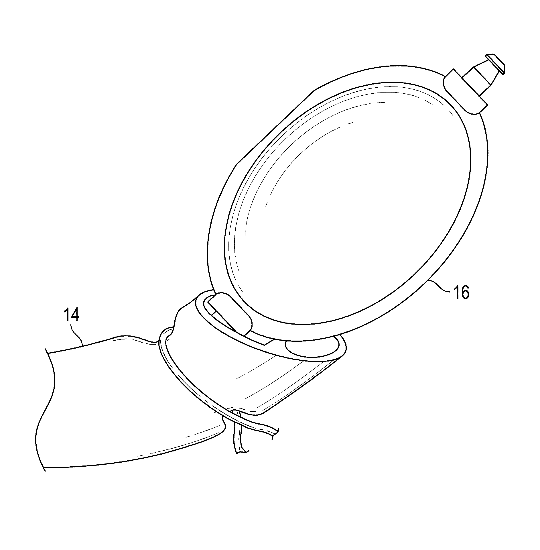
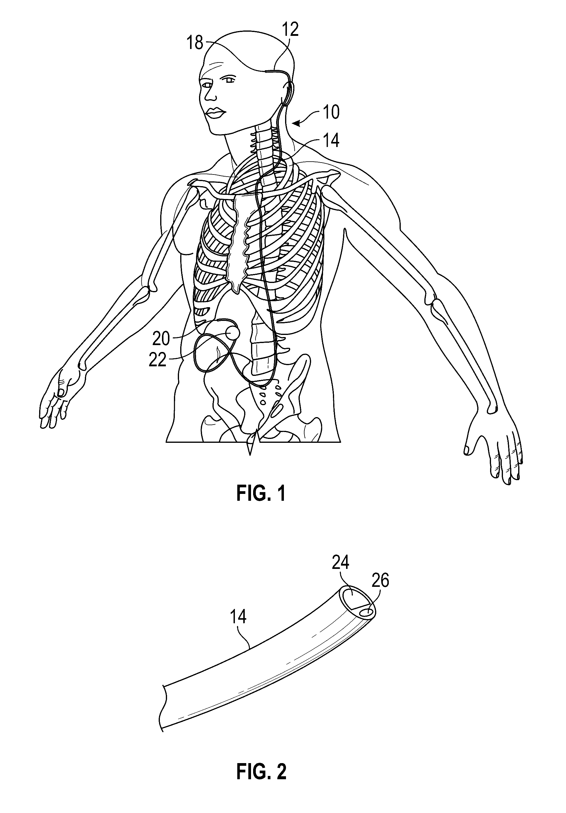
[0026]The ventriculoperitoneal shunt 10 of the invention includes a proximal or ventricular catheter 12 and a distal or peritoneal catheter 14. A shunt valve 16 connects the adjacent ends of the catheters 12, 14. The proximal or free end 18 of the ventricular catheter 12 is adapted to be inserted into the brain ventricle, while the distal or free end 20 of the peritoneal catheter 14 is adapted to be inserted into the peritoneal cavity, with the catheter 12 tunneling beneath the skin between the opposite ends 18, 20. The catheters, when connected to the valve 16, form a continuous catheter drain path from the brain ventricle to the peritoneal cavity.

[0027]The above described structure for the VP shunt 10 is conventional.

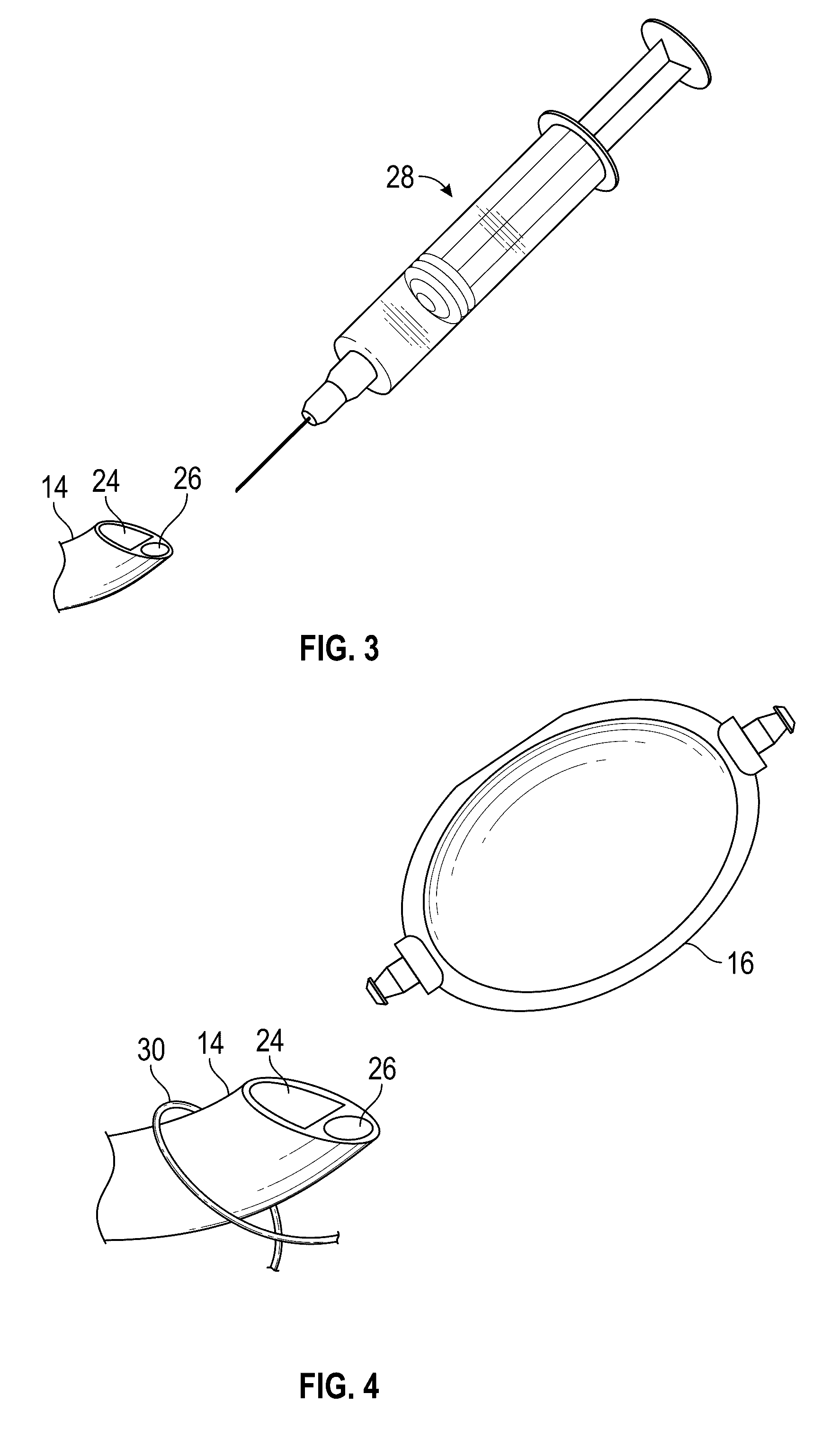
[0028]The present invention improves the conventional VP shunt 10 by incorporating an inflatable balloon 22 on the distal end of the catheter 14. The balloon resides within the peritoneal cavity, and is surgically implanted in a deflated condition. During surgery, the...


PUM


Abstract

A ventriculoperitoneal shunt has a ventricular catheter and a peritoneal catheter, with a shunt valve connected in line between the catheters to define a drainage patch for cerebrospinal fluid from the brain ventricle to the peritoneal cavity. An inflatable balloon on the peritoneal catheter precludes migration or displacement of the catheter in the peritoneal cavity, thereby eliminating or minimizing corrective revision surgery.

Description

CROSS REFERENCE TO RELATED APPLICATIONS[0001]This application claims priority under 35 U.S.C. §119 to provisional application U.S. Ser. No. 62 / 144,496 filed Apr. 8, 2015, all of which is herein incorporated by reference in its entirety.BACKGROUND OF THE INVENTIONS[0002]Certain medical conditions, such as head injuries, congenital malformation, bleeding from a blood vessel in the brain, idiopathic intracranial hypertension and meningitis, can produce increase in intracranial pressure (ICP) by preventing normal drainage of cerebrospinal fluid (CSF). Such CSF buildup typically occurs in the ventricles inside the brain, which cause the ventricles to enlarge and compress the brain against the skull, leading to increased ICP. This increase in ICP can cause serious neurological problems, including death. This condition is known medically as hydrocephalus.[0003]In conditions where the cerebrospinal fluid must be drained for significant periods of time, a ventriculoperitoneal (VP) shunt is t...

Claims


Application Information

Patent Timeline
Patent Type & Authority Applications(United States)
IPC IPC(8): A61M27/00
CPCA61M2025/1079A61M27/006
Inventor FLOUTY, OLIVER ELIASABODE-IYAMAH, KINGSLEY OTSIORENHOWARD, MATTHEW A.GREENLEE, JEREMY D.
Owner UNIV OF IOWA RES FOUND
Why Eureka
Try Eureka