Methods, systems, and computer programs for segmenting a tooth's pulp region from an image

a technology of tooth pulp and image, applied in image enhancement, medical/anatomical pattern recognition, instruments, etc., can solve the problems of pulp death, jaw bone loss, tooth loss, etc., and achieve the effect of convenient and precise planning of endodontic treatmen

Active Publication Date: 2021-07-08
MEDICIM
View PDF0 Cites 2 Cited by
  • Summary
  • Abstract
  • Description
  • Claims
  • Application Information

AI Technical Summary

Benefits of technology

[0011]This enables the efficient and automatic segmentation of a pulp region from 2D or 3D image data starting from a (e.g., user-selected) seed point in the pulp chamber. In particular, an iterative process enables the segmentation of the pulp region. In the iterative process, the grayscale thresholding (GT) aims at detecting the pulp region based on intensity values of image elements, whereas the curvature-based shape recognition (CBSR) applied to the image elements complements and combines with the GT to detect typical structures of root canals, such as tubular and sheet-like shapes, while discarding parts unlikely to constitute root canals. Further, the method is well suited to enable, in at least some embodiments, the detection of potentially intricate shapes of certain root canals, including bifurcations and obstructions therein, so as to eventually enable the convenient and precise planning of an endodontic treatment.

Problems solved by technology

When a tooth is cracked or has a deep cavity, bacteria may enter the pulp and cause serious infections.
When untreated, such infections of the root canal may result in pulp death, jaw bone loss and loss of the tooth itself (see ref.
A difficulty during root canal treatment is cleaning and filling all root canals over their entire length.
If parts are missed, infections are likely to occur afterwards.
This may potentially cause serious pain and inflammations for the patients.
Some teeth have complex canal shapes that are hard to visualize using a 2D or even a 3D image.
Although the method of Jud et al. allows predicting the probability that an image element is within the pulp region, the authors indicate that the high variability in between teeth of the shape of the pulp region is incongruent with building a statistical pulp shape model and registering such model to pulp object probabilities.
As such the method of Jud et al. cannot be used for segmenting the pulp region of a tooth.

Method used


Image

  • Methods, systems, and computer programs for segmenting a tooth's pulp region from an image
  • Methods, systems, and computer programs for segmenting a tooth's pulp region from an image
  • Methods, systems, and computer programs for segmenting a tooth's pulp region from an image

Examples


Embodiment Construction

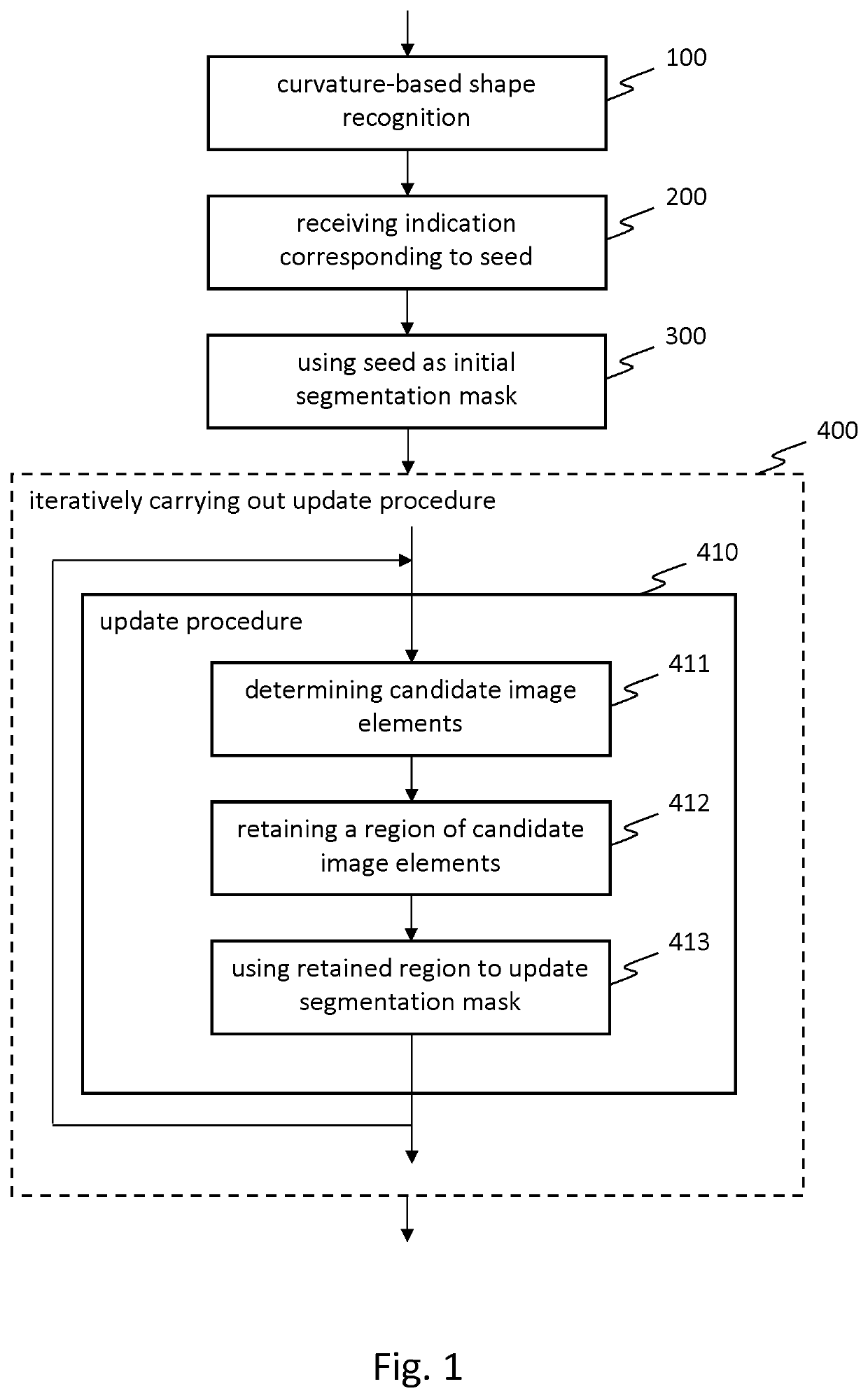
[0032]The present development shall now be described in conjunction with specific embodiments. These specific embodiments serve to provide the skilled person with a better understanding, but are not intended to in any way restrict the scope of the development, which is defined by the appended claims. A list of abbreviations and their meaning is provided at the end of the detailed description.

[0033]FIG. 1 is a flowchart of a method in one embodiment of the development. The method is computer-implemented. That is, the method may be carried out by a computer or set of computers, although the development is not limited thereto. The method may also be implemented, as a form of computer-implemented method, using hardware electronic circuits, e.g. with field-programmable gate arrays (FPGA), or using a combination of computer(s) and hardware electronic circuits.

[0034]The method aims at segmenting a tooth's pulp region from a 2D or 3D image of said tooth. The pulp region comprises a pulp cha...


PUM


Abstract

Methods, systems, and computer programs are disclosed for segmenting, from an image, a tooth's pulp region comprising a pulp chamber and root canals. Curvature-based shape recognition is performed at different spatial scales using smoothed versions of the image. An indication of a point or region is received, located in the pulp chamber and referred to as “seed”. The seed is used as initial segmentation mask. An update procedure, iteratively carried out, comprises: (i) determining candidate image elements for updating the segmentation mask, comprising: (i−1) in the first n iteration(s), a grayscale thresholding taking as reference the current segmentation mask; and, (i−2) in at least one iteration, taking the curvature-based shape recognition into account; (ii) retaining, among the candidate image elements, a region of connected candidate image elements that comprises the seed; and (iii) using the retained region to update the segmentation mask.

Description

INCORPORATION BY REFERENCE TO ANY PRIORITY APPLICATIONS[0001]Any and all applications for which a foreign or domestic priority claim is identified in the Application Data Sheet as filed with the present application are hereby incorporated by reference under 37 CFR 1.57.[0002]This application is filed under 35 U.S.C. § 371 as the U.S. National Phase of Application No. PCT / EP2019 / 063883 entitled “METHODS, SYSTEMS, AND COMPUTER PROGRAMS FOR SEGMENTING A TOOTH'S PULP REGION FROM AN IMAGE” and filed May 28, 2019, and which claims priority to EP 18174919.3 filed May 29, 2018, each of which is incorporated by reference in its entirety.BACKGROUND OF THE INVENTIONField of the Invention[0003]The present development relates to methods for segmenting a tooth's pulp region from a two-dimensional (2D) or three-dimensional (3D) image of said tooth. The development also relates to systems for carrying out such methods, and to computer programs therefor. Some embodiments of the development may for e...

Claims


Application Information

Patent Timeline
Patent Type & Authority Applications(United States)
IPC IPC(8): G06T7/11G06K9/32G06K9/46G06T7/50G06T7/00G06T7/136G06T7/194A61B6/14
CPCG06T7/11G06K9/3233G06K9/4604G06T7/50G06T7/0014G06K2209/05G06T7/194A61B6/14G06T2207/30036G06T2207/10116G06T7/136G06T7/187G06T2207/10081G06T2207/20101G06T2207/20164A61B6/032A61B6/4085G06V2201/03
Inventor GORIS, BARTVAN LEEMPUT, PIETERMOLLEMANS, WOUTERELEN, AN
Owner MEDICIM
Why Eureka
Try Eureka