Method for simultaneous body part display

a body part and display method technology, applied in the field of simultaneous display of breasts obtained using mammography equipment, to achieve the effect of simple and practical comparison

Inactive Publication Date: 2006-05-16
GE MEDICAL SYST GLOBAL TECH CO LLC
View PDF9 Cites 15 Cited by
  • Summary
  • Abstract
  • Description
  • Claims
  • Application Information

AI Technical Summary

Benefits of technology

[0005]There is consequently a need for a type of display providing simple and practical comparison of the two images obtained in examination apparatus. This problem is particularly prevalent for images of both breasts obtained in a mammograph.
[0006]Briefly, in one embodiment of the present inventio

Problems solved by technology

This problem is particularly prevalent for im

Method used


Image

  • Method for simultaneous body part display
  • Method for simultaneous body part display
  • Method for simultaneous body part display

Examples


Embodiment Construction

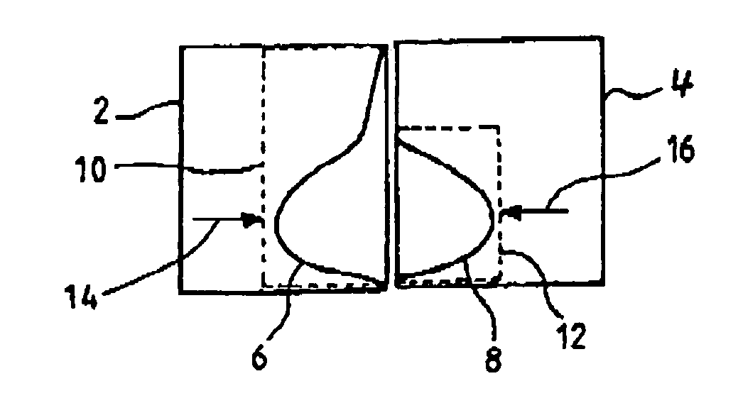
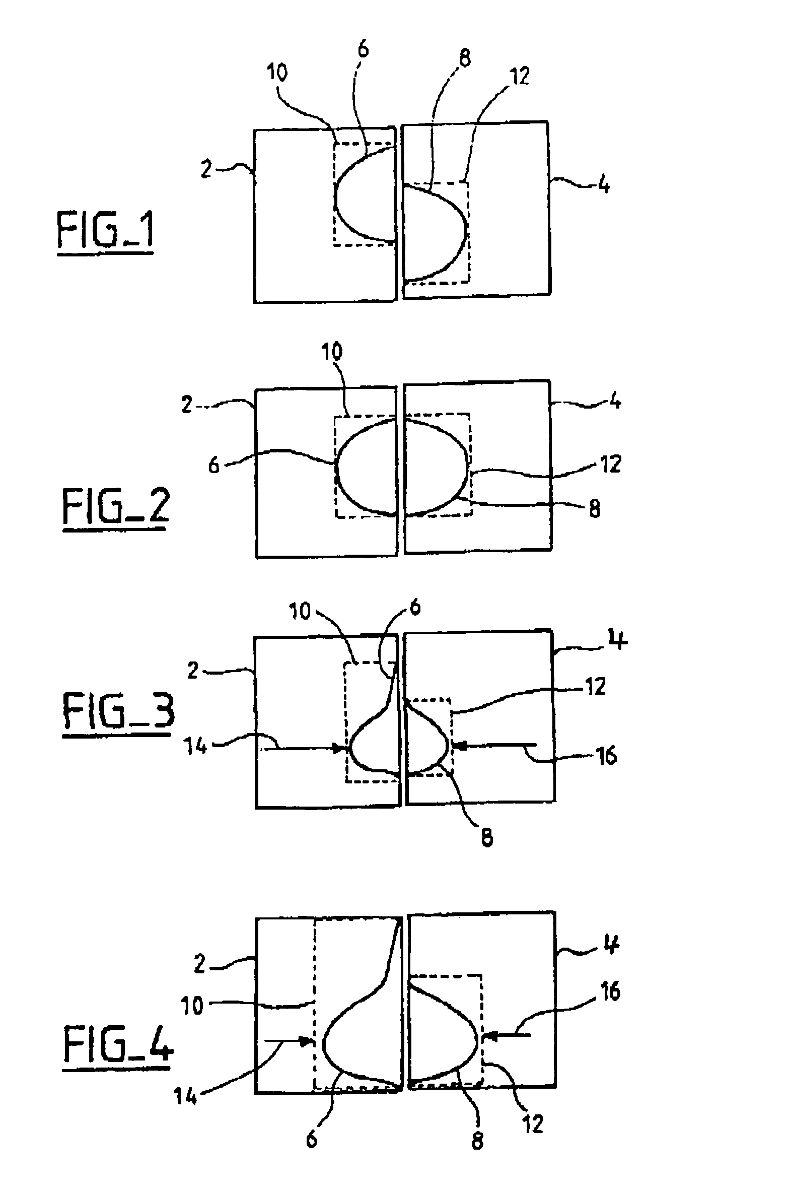
[0013]The embodiments of the invention will be described with reference to its preferred application to the display of digital images of both breasts supplied by mammography apparatus. These images can be images acquired digitally for subsequent digital processing for their display. The images can also be obtained from analog radiography apparatus by exposure and development of films. The analog images can then be digitized and displayed as discussed below.

[0014]FIG. 1 shows diagrammatically images 2 and 4 of both breasts displayed in a known apparatus. The first image 2 shows the right-hand breast of a patient in a cranio-caudal view. The second image 4 shows the patient's left-hand breast in a cranio-caudal view. Both images are displayed side by side, the first image being to the left of the second image. The respective contours 6 and 8 of the breasts are shown diagrammatically on their images 2 and 4. On the images 2 and 4, regions of interest 10 and 12 defined around the breast...


PUM


Abstract

The regions of interest of images of both breasts of a patient are defined as regions of the image of minimal surface area and containing views of the breasts. These regions of interest are aligned for simultaneous display of the images. The display now allows simple and direct comparison of the images of both breasts, notably allowing immediate comparison of structural disorganizations within the two breasts.

Description

CROSS REFERENCE TO RELATED APPLICATIONS[0001]This application claims the benefit of a priority under 35 USC 119 to French Patent Application No. 00 16 957 filed Dec. 22, 2000, the entire contents of which are incorporated by reference.FIELD OF THE INVENTION[0002]The invention relates to the display of digital images of body parts and more particularly to simultaneous display of the breasts of a patient obtained using mammography apparatus.BACKGROUND OF THE INVENTION[0003]Mammography apparatus is used for examining patients' breasts using X-rays. The image is acquired using an image receiver formed either by a photographic plate or by digital sensing means. Examinations practiced with such apparatus comprise taking cranio-caudal and lateral images Cranio-caudal views are obtained by irradiating the breast from above so as to obtain a view in the axis running from the patient's head to her feet. To obtain a side view, the breast is irradiated from the side so as to obtain a view in an...

Claims


Application Information

Patent Timeline
IPC IPC(8): G06K9/32A61B6/02G06K9/00G06T5/00G06T7/00
CPCG06T7/0012G06T7/0024G06T2207/30068G06T2207/20132G06T2207/10116G06T7/30
Inventor SOUBELET, ELISABETHBOTHOREL, SYLVIEMULLER, SERGE LOUIS
Owner GE MEDICAL SYST GLOBAL TECH CO LLC
Why Eureka
Try Eureka