Magnetic resonance tomography method and apparatus with motion correction

a technology of magnetic resonance tomography and motion correction, which is applied in the direction of magnetic variable regulation, instruments, applications, etc., can solve the problems of no longer being reliable whether, perforation of the vessel wall, and no longer being able to achieve the effect of reducing the risk of vascular damag

Inactive Publication Date: 2007-05-22
SIEMENS HEALTHCARE GMBH
View PDF4 Cites 35 Cited by
  • Summary
  • Abstract
  • Description
  • Claims
  • Application Information

AI Technical Summary

Benefits of technology

[0008]It is an object of the present invention to provide a magnetic resonance tomography apparatus and a method for the implementation of magnetic resonance tomography which allow a motion correction of the vessel image to be implemented in an angiography procedure with magnetic resonance-monitored vessel intervention, and to make an image available of the medical intervention device that corresponds to the true position with reference to the vessel system.
[0013]Using a technique already known in the framework of other medical examinations, the respiratory motion of the patient can be advantageously acquired in a simple and dependable way, both with respect to its momentary position and its maximum expanse and motion.
[0015]It is thus possible to acquire the coarse of respiration of the patient without further auxiliary devices at the patient.
[0019]With such an apparatus a fluid and a small, narrowly bounded section can be acquired well and quickly in magnetic resonance tomography. As a result, it is possible to reconstruct the motion of the entire vessel system from the motion of this small section. Particularly when a number of such sections are defined, the motion of the vessel system can be identified relatively precisely. Thus, an attending physician can implement an angiography examination with vessel interventions with magnetic resonance tomography just with the previously described magnetic resonance tomography and can monitor the exact position of a catheter.
[0021]Such a slice excitation perpendicular to a larger vessel such as, for example, the aorta descendens, arteria iliaca communis, or the arteria renalis can be easily accomplished with known methods of magnetic resonance tomography. In particular, the acquisition of such a follow-up section, since it need ensue only with less detail and less highly resolved and is only intended to acquire the motion sequence, can ensue very quickly. It can thus also easily track the motion.
[0023]As a result, it is possible for the correction of the road map to be precise and exact in the vessel section that is particularly important for the medical diagnostic in the present treatment method. Because a correction only ensues when the intervention device, i.e. particularly a catheter, has apparently penetrated a vessel wall, a check can be made within the magnetic resonance tomography apparatus with little calculating outlay and expense to determine whether this has truly occurred. Further, the vessel section wherein the end of the catheter is located can be detected and found especially well, since a striking contrast in the image can be located with the end of the catheter and it is consequently only necessary to undertake a further registration of the tissue and of the liquids in the region around this end segment.

Problems solved by technology

The problem can occur that the first exposure, the road map, no longer corresponds to the current condition and position of the vessel system, since modifications and distortions of the vessel system occur both due to gross movements on the part of the patient but primarily due to respiratory motion.
It can then no longer be reliable whether the medical examination device is still located within a vessel or whether the vessel wall has been perforated, since the catheter and / or guide wire at least sporadically produce the impression in the superimposed exposure that they are located outside the vessel tree.
Such a perforation of the vessel wall is a complication that is definitely possible and must be taken very seriously in an interventional vessel operation.
The described problem occurs not only when probing or catheterizing smaller vessels wherein the vessel diameter lies on the order of magnitude of the medical intervention device (for example, catheter or guide wire) but also occurs in interventions in larger vessels, since the medical intervention devices do not necessarily lie in the center of the vessel cross-section, but have the tendency to conform to the vessel wall and to be guided thereby when advanced farther into the vessel.

Method used


Image

  • Magnetic resonance tomography method and apparatus with motion correction
  • Magnetic resonance tomography method and apparatus with motion correction
  • Magnetic resonance tomography method and apparatus with motion correction

Examples


Embodiment Construction

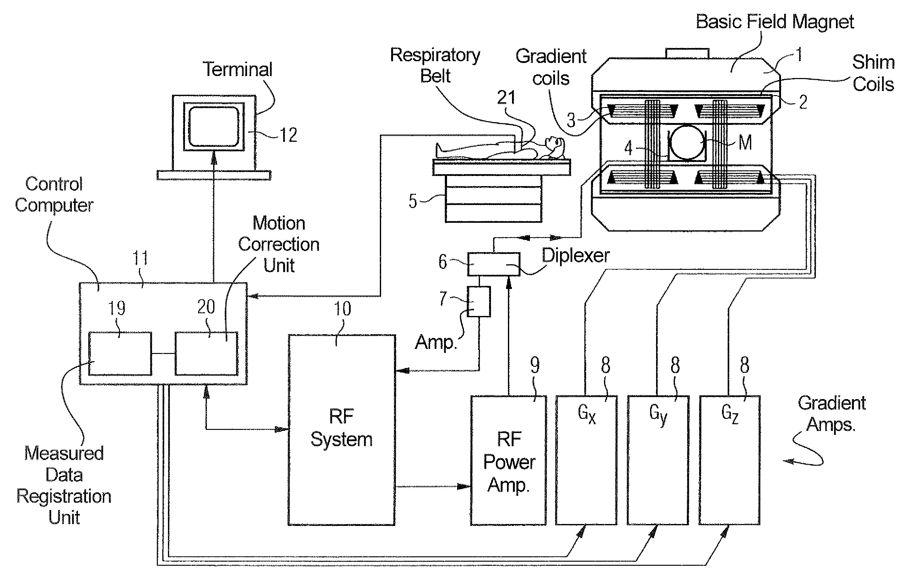
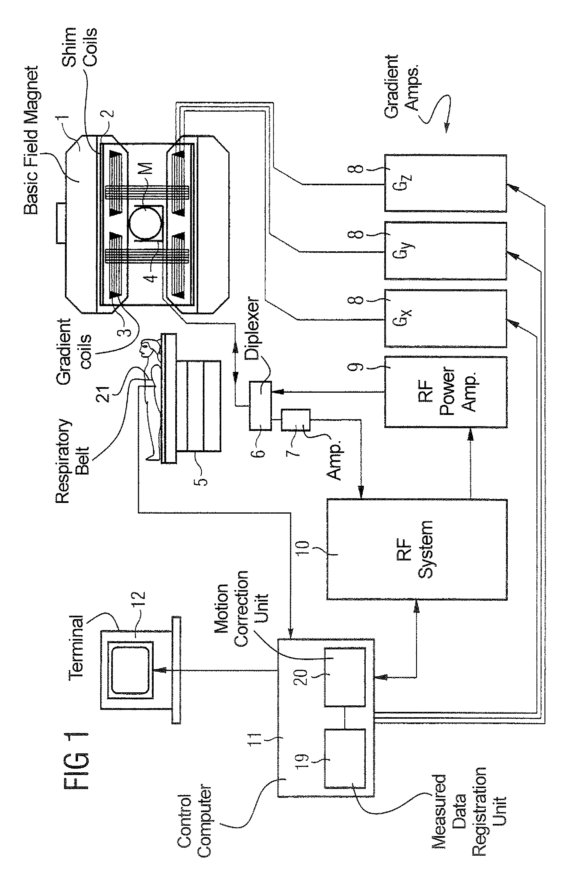
[0034]In a schematic illustration, FIG. 1 shows the structure of a magnetic resonance tomography apparatus that corresponds to the structure of a conventional magnetic resonance tomography apparatus but is operable according to the invention. A basic field magnet 1 generates a strong, optimally uniformly magnetic field for polarization of the nuclear spins in an examination region in the inside of the basic field magnet 1. The high uniformity of the basic magnet field required for a magnetic resonance measurement is mainly defined in a spherical measurement volume M. On a movable supporting table 5, a person can be introduced into the inside of the basic field magnet 1 so that the region of the human body that is to be examined is located in the inside of the measurement volume M. For correcting time-invariable influences, shim plates of ferromagnetic material are attached to suitable locations in addition to the basic field magnet 1. Further, a correction for time-variable influenc...


PUM


Abstract

In a magnetic resonance tomography apparatus and method with motion correction in an angiography examination with magnetic resonance-monitored vessel intervention, a contrast agent-supported exposure of the vessel system (road map) is registered, with one exposure ensuing at the end of expiration and one exposure ensuing at the end of inspiration of the patient, and a magnetic resonance-visible medical intervention device introduced into the vessel system also is registered. A corrected road map of the vessel system is interpolated from the exposures at the end of inspiration and the end of expiration and the exposure of the intervention device is superimposed on the corrected road map.

Description

BACKGROUND OF THE INVENTION[0001]1. Field of the Invention[0002]The present invention is directed to a magnetic resonance tomography apparatus of the type having a device for motion correction and to a method for motion correction in a magnetic resonance exposure in an angiography procedure with magnetic resonance-monitored vessel intervention. In particular, the present invention is directed to a magnetic resonance tomography apparatus and method wherein, an angiography procedure, a medical intervention device can be introduced into the vessel system and monitored with by magnetic resonance as to its location within the vessel system, and to thereby corrects for movements of the patient such as respiratory motion.[0003]2. Description of the Prior Art[0004]As a medical diagnostic modality, nuclear magnetic resonance tomography has also proven well-suited in order to implement an angiography procedure, a registration of a vessel system, and, in particular, to monitor the introduction...

Claims


Application Information

Patent Timeline
Patent Type & Authority Patents(United States)
IPC IPC(8): A61B5/055G01R33/28G01R33/563G01R33/567
CPCG01R33/5673G01R33/5676G01R33/5635A61B5/055
Inventor NITZ, WOLFGANG
Owner SIEMENS HEALTHCARE GMBH
Why Eureka
Try Eureka