Vessel segmentation with nodule detection

Inactive Publication Date: 2007-09-18
MEVIS MEDICAL SOLUTIONS
View PDF7 Cites 21 Cited by
  • Summary
  • Abstract
  • Description
  • Claims
  • Application Information

AI Technical Summary

Benefits of technology

[0018]The present invention is a system for efficiently detecting and highlighting anatomical information obtained from a digital acquisition system. In particular, seeds are identified and grown into regions in a controlled manner. The regions processed to highlight potentially cancerous nodules and other particles or objects.

Problems solved by technology
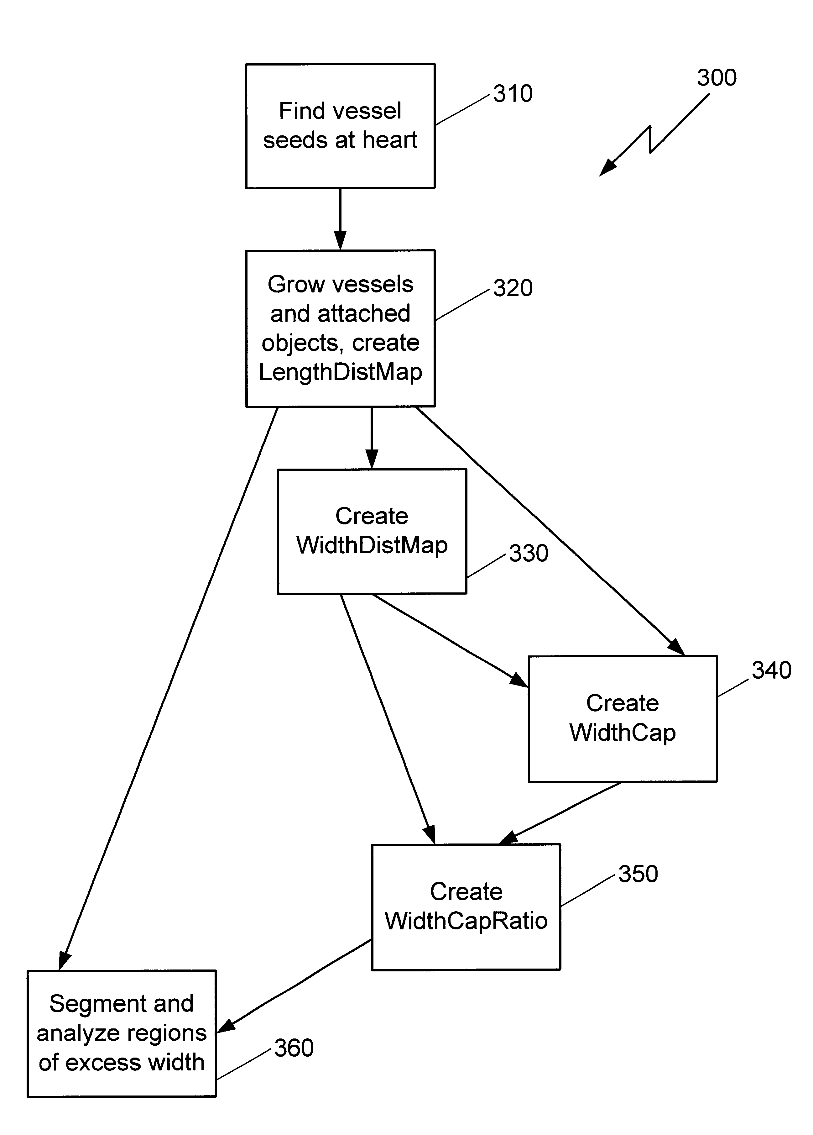
Unfortunately, in all too many cases, this application is merely to confirm the worst.
Region growing techniques, as opposed to edge-based segmentation techniques, are particularly effective in noisy images since edges in noisy images are extremely difficult to detect.
Key issues in digital image processing are speed and accuracy.

Method used


Image

  • Vessel segmentation with nodule detection
  • Vessel segmentation with nodule detection
  • Vessel segmentation with nodule detection

Examples


Embodiment Construction

[0033]Modern CT scanners, particularly multidetector scanners which acquire more than one slice per gantry rotation, provide structural data of unprecedented quality due to their favorable volumetric resolution and acquisition speed. This data has superior diagnostic value, enabling the detection of potential cancers or other health problems at early and more treatable stages. Similarly, detecting a particle or obstacle that can restrict the flow of blood is helpful in assessing the health of a patient regarding the adequate flow of blood to vital organs.

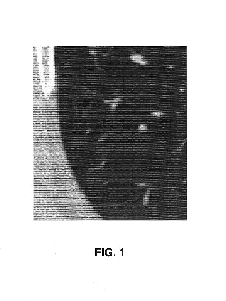
[0034]Within various images of the anatomy, such as images including the lungs or the heart or portions thereof, numerous grey flecks may appear on a CT axial scan or other digital image. For the most part, these flecks may be sections of blood vessels that have absorbed sufficient x-rays to be visible in the grey scale image. FIG. 1 depicts a digital axial section of a thoracic region with various visible cross-sections of blood ve...


PUM


Abstract

A region-growing method for identifying nodules in an anatomical volume segments a 3-D image volume by controlled voxel growth from seed points. The process is based on creation and use of a distance map for tracking the distance of vessel voxels from a predetermined location. A volume map is created that identifies the largest sphere that can pass between a voxel and a predetermined location without touching a non-vessel voxel. The ratio between the distance map and the volume map is analyzed to find regions more likely to contain nodules, the features of which can be extracted or otherwise highlighted.

Description

CROSS-REFERENCE TO RELATED APPLICATIONS[0001]Related applications are:[0002]“Density Nodule Detection in 3-Dimensional Medical Images,” Ser. No. 09 / 993,792, filed concurrently herewith;[0003]“Method and System for the Display of Regions of Interest in Medical Images,” Ser. No. 09 / 990,508, filed Nov. 21, 2001, attorney docket number 8498-039-999;[0004]“Automated Registration of 3-D Medical Scans of Similar Anatomical Structures,” Ser. No. 09 / 993,790, filed concurrently herewith;[0005]“Lung Field Segmentation From CT Thoracic Images,” Ser. No. 09 / 993,793, filed concurrently herewith;[0006]“Pleural Nodule Detection from CT Thoracic Images,” Ser. No. 09 / 993,789, filed concurrently herewith; and[0007]“Graphical User Interface for Display of Anatomical Information,” Ser. No. 09 / 990,511, filed Nov. 21, 2001, claiming priority from Ser. No. 60 / 252,743, filed Nov. 22, 2000 and from Ser. No. 60 / 314,582 filed Aug. 24, 2001.[0008]This application hereby incorporates by reference the entire disc...

Claims


Application Information

Patent Timeline
IPC IPC(8): G06K9/00A61B6/03G06F3/033G06F3/048G06F19/00G06T5/00G06T7/00G06T11/00G06T15/08G06V10/25
CPCA61B6/032A61B6/50G06F3/0481G06F3/04855G06F19/321G06F19/3406G06F19/3437G06K9/3233G06K9/342G06T7/0012G06T7/0081G06T15/08A61B6/463G06T2207/30101G06F19/363G06F2203/04805G06K2009/00932G06K2009/366G06K2209/05G06T2207/10081G06T2207/20101G06T2207/20141G06T2207/30048G06T2207/30061G06T7/11G06T7/187G16H10/20G16H40/63G16H50/50G16H30/40G06V40/14G06V10/248G06V10/25G06V10/267G06V2201/03
Inventor SCHNEIDER, ALEXANDER C.WOOD, SUSAN A.
Owner MEVIS MEDICAL SOLUTIONS
Why Eureka
Try Eureka