Region growing in anatomical images
a technology of anatomical images and regions, applied in image enhancement, medical/anatomical pattern recognition, instruments, etc., can solve the problems of speed and accuracy of processing, and achieve the effect of efficient detection and highlighting
- Summary
- Abstract
- Description
- Claims
- Application Information
AI Technical Summary
Benefits of technology
Problems solved by technology
Method used
Image
Examples
Embodiment Construction
[0038]Modern CT scanners, particularly multidetector scanners which acquire more than one slice per gantry rotation, provide structural data of unprecedented quality due to their favorable volumetric resolution and acquisition speed. This data has superior diagnostic value, enabling the detection of potential cancers or other health problems at early and more treatable stages. Similarly, detecting a particle or obstacle that can restrict the flow of blood is helpful in assessing the health of a patient regarding the adequate flow of blood to vital organs.
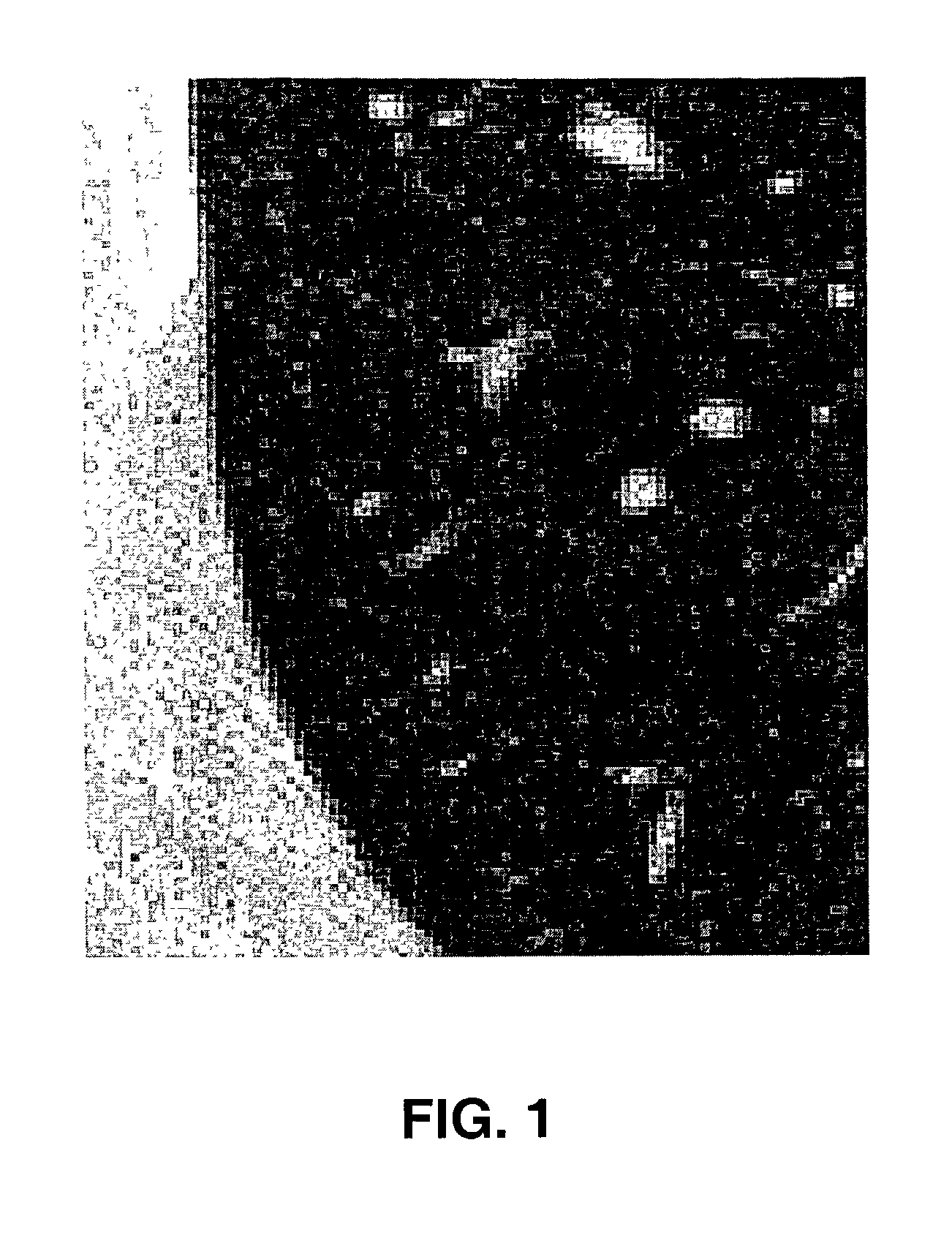
[0039]Within various images of the anatomy, such as images including the lungs or the heart or portions thereof, numerous grey flecks may appear on a CT axial scan or other digital image. For the most part, these flecks may be sections of blood vessels that have absorbed sufficient x-rays to be visible in the grey scale image. FIG. 1 depicts a digital axial section of a thoracic region with various visible cross-sections of blood ve...
PUM
 Abstract
Description
Claims
Application Information
 - R&D
- Intellectual Property
- Life Sciences
- Materials
- Tech Scout
- Unparalleled Data Quality
- Higher Quality Content
- 60% Fewer Hallucinations
Browse by: Latest US Patents, China's latest patents, Technical Efficacy Thesaurus, Application Domain, Technology Topic, Popular Technical Reports.
© 2025 PatSnap. All rights reserved.Legal|Privacy policy|Modern Slavery Act Transparency Statement|Sitemap|About US| Contact US: help@patsnap.com


