Method and apparatus for taking a biopsy

a biopsy and endoscope technology, applied in the field of medicine, can solve the problems of inability to directly visualize intraventricular mass lesions, surgeons are reluctant to biopsy intraventricular mass lesions, and intraventricular mass lesions, on the other hand, provide no structural suppor

Inactive Publication Date: 2008-02-26
RGT UNIV OF MINNESOTA
View PDF14 Cites 229 Cited by
  • Summary
  • Abstract
  • Description
  • Claims
  • Application Information

AI Technical Summary

Problems solved by technology

Because of this requirement, endoscopy devices are limited in their applications.
In addition, surgeons are occasionally reluctant to biopsy intraventricular mass lesions without direct visualization because of the risk of hemorrhage.
Intraventricular lesions, on the other hand, provide no such structural support as a barrier to postbiopsy hemorrhage.

Method used


Image

  • Method and apparatus for taking a biopsy
  • Method and apparatus for taking a biopsy
  • Method and apparatus for taking a biopsy

Examples


Embodiment Construction

[0007]In the following detailed description of the invention reference is made to the accompanying drawings which form a part hereof, and in which is shown, by way of illustration, specific embodiments in which the invention may be practiced. In the drawings, like numerals describe substantially similar components throughout the several views. These embodiments are described in sufficient detail to enable those skilled in the art to practice the invention. Other embodiments may be utilized and structural, logical, and electrical changes may be made without departing from the scope of the present invention.

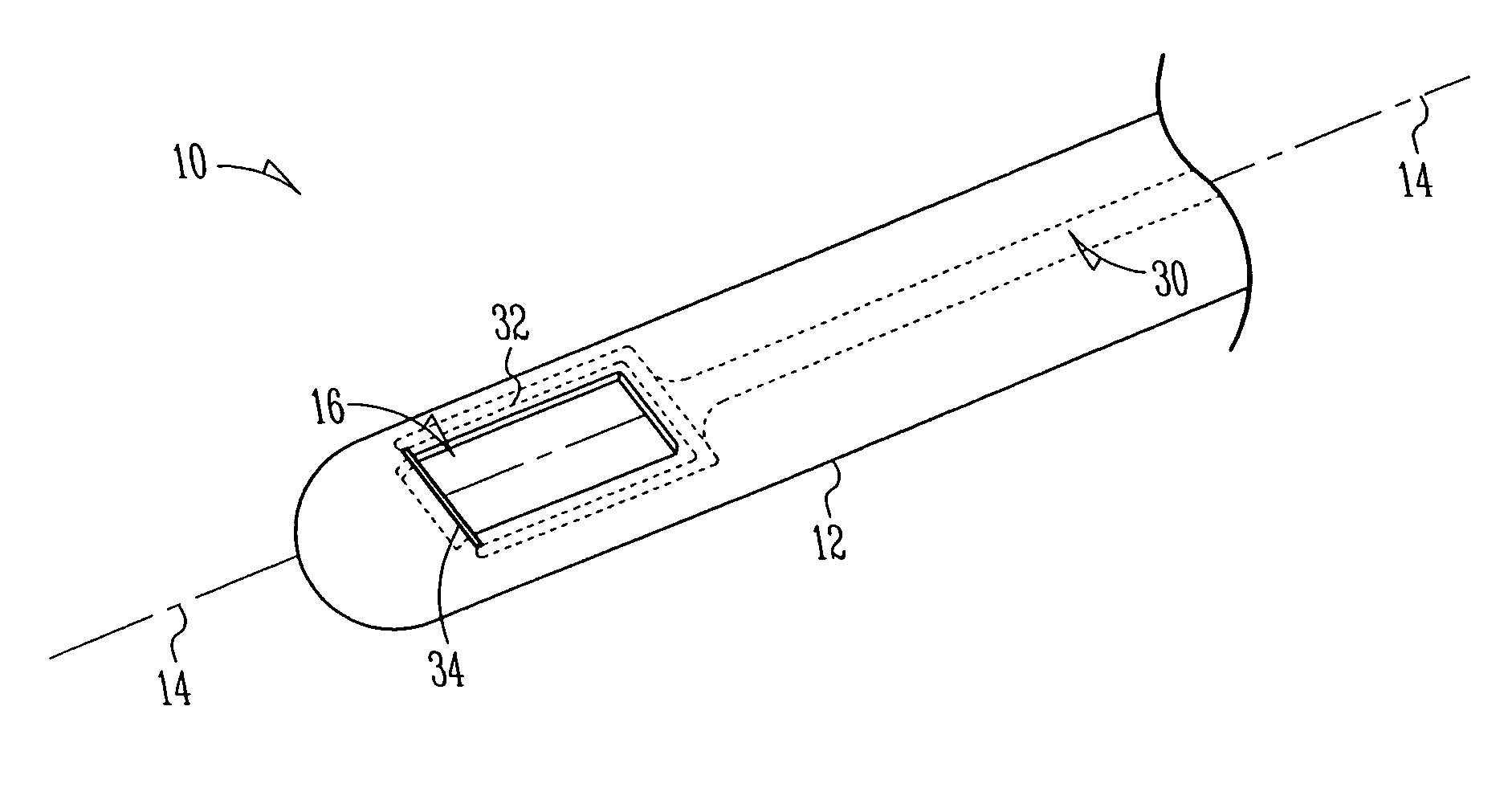
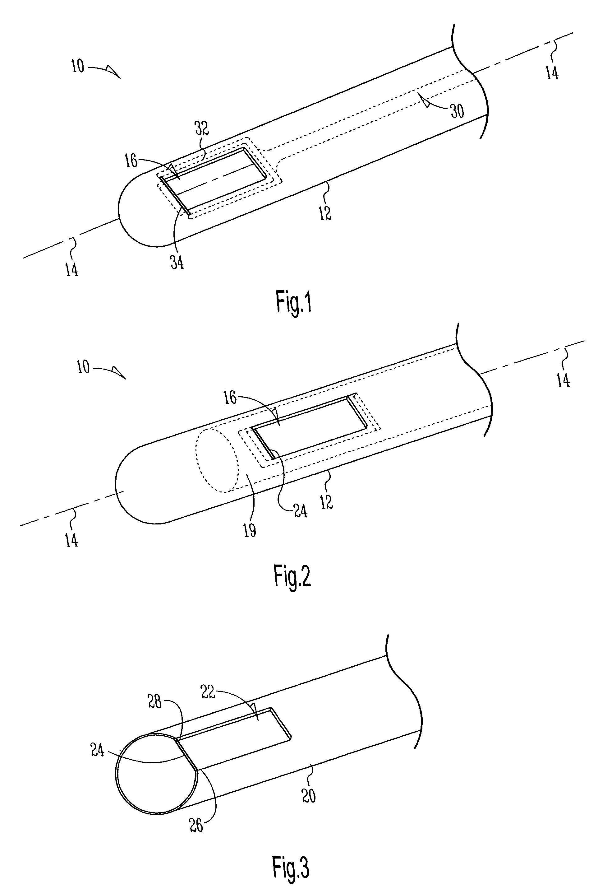
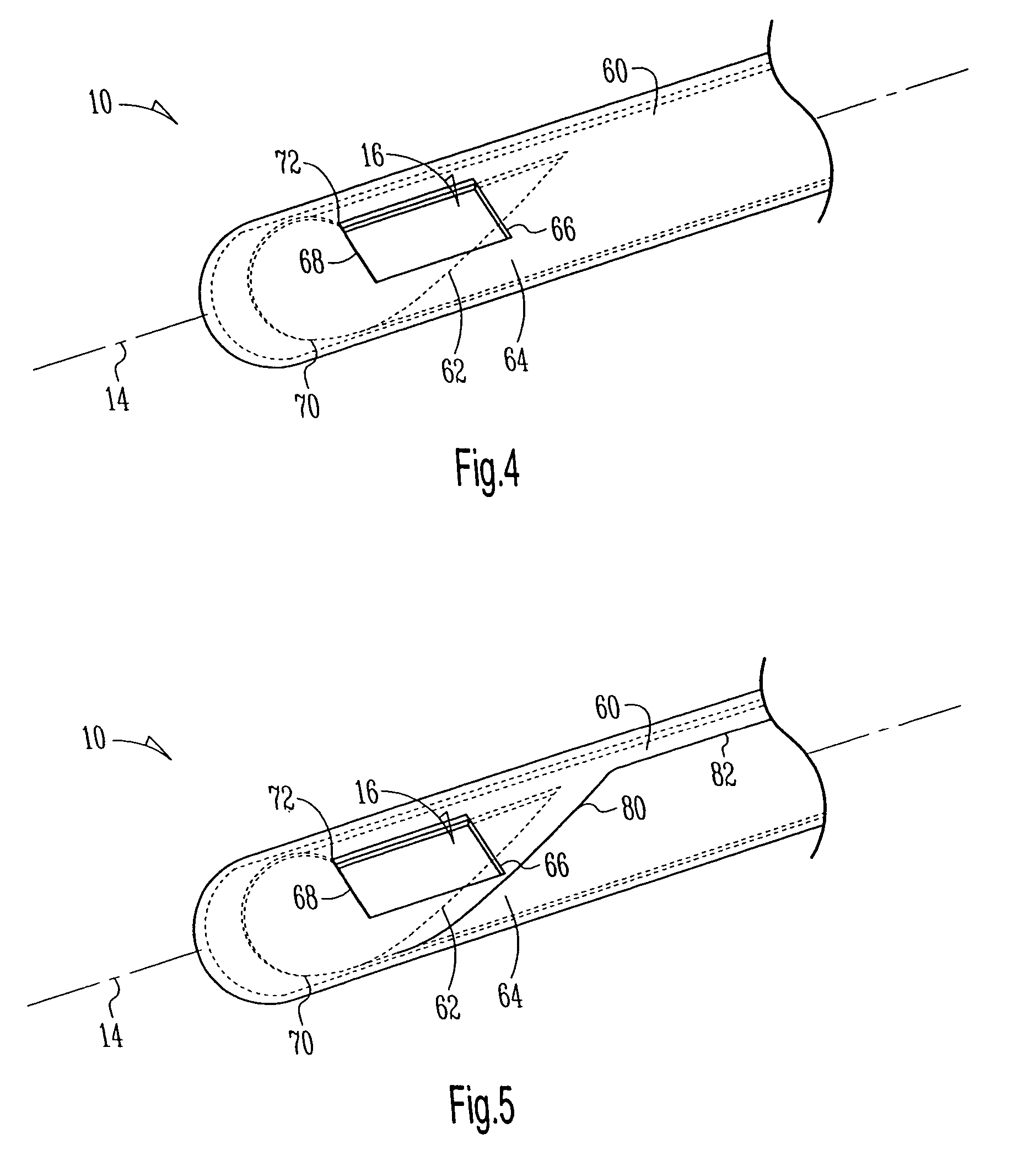
[0008]Referring now to FIG. 1, there is illustrated an example embodiment of method and apparatus for taking a biopsy. An introducer 10 is formed of a biocompatible cylindrical member 12 with an outer wall and a hollow core, the cylindrical member 12 having a longitudinal axis 14. An opening 16 in the outer wall of the member is provided wherein the opening extends, in one example ...


PUM


Abstract

There is described method and apparatus for taking a biopsy wherein the apparatus is formed of a biocompatible cylindrical member with an outer wall and a hollow core, the cylindrical member having a longitudinal axis. An opening in the outer wall of the member is provided and a cutting device cuts tissue entering the opening as it travels along the opening. Cut tissue is treated with a coagulating agent or member so that the coagulation occurs concurrently or just after the tissue is cut wherein the coagulation occurs at a trailing edge of the opening as it travels.

Description

RELATED APPLICATIONS[0001]This application is a continuation of U.S. patent application Ser. No. 09 / 919,583, filed on Jul. 31, 2001 now U.S. Pat. No. 6,773,443 which claims priority under 35 U.S.C. § 119(e) of U.S. Provisional Application No. 60 / 221,712, filed Jul. 31, 2000, which applications are incorporated herein by reference.TECHNICAL FIELD OF THE INVENTION[0002]The present invention relates generally to the field of medicine, and more particularly to an endoscopy biopsy device and its use in medical procedures.BACKGROUND[0003]Intracranial endoscopy devices include both flexible and rigid endoscopes. These devices typically have at least one channel through which a biopsy device or grabbing forcep device can be delivered. As a result, the combination of a fiberoptic endoscopic channel with a second channel for biopsy device necessitates a minimum diameter. Because of this requirement, endoscopy devices are limited in their applications. Specifically, some neurosurgeons prefer n...

Claims


Application Information

Patent Timeline
Patent Type & Authority Patents(United States)
IPC IPC(8): A61B17/32A61B10/00A61B10/04A61B17/22A61B18/14A61B19/00
CPCA61B10/04A61B17/320068A61B17/320783A61B18/14A61B2017/00911A61B2017/320069A61B2017/32007
Inventor TRUWIT, CHARLES L.NELSON, RANDAL
Owner RGT UNIV OF MINNESOTA
Why Eureka
Try Eureka