Method for recording projection image

a projection image and image technology, applied in medical science, diagnostics, electrical equipment, etc., can solve the problem of only being able to optimize the recording parameters, and achieve the effect of minimizing radiation exposure, maximizing the image quality of the projection image, and avoiding radiation exposure of the object being examined

Inactive Publication Date: 2008-05-13
SIEMENS AG
View PDF3 Cites 6 Cited by
  • Summary
  • Abstract
  • Description
  • Claims
  • Application Information

AI Technical Summary

Benefits of technology

[0007]Proceeding from this prior art, the object of the invention is therefore to disclose an adaptive method for recording projection images for minimizing the exposure to radiation of the object being examined.
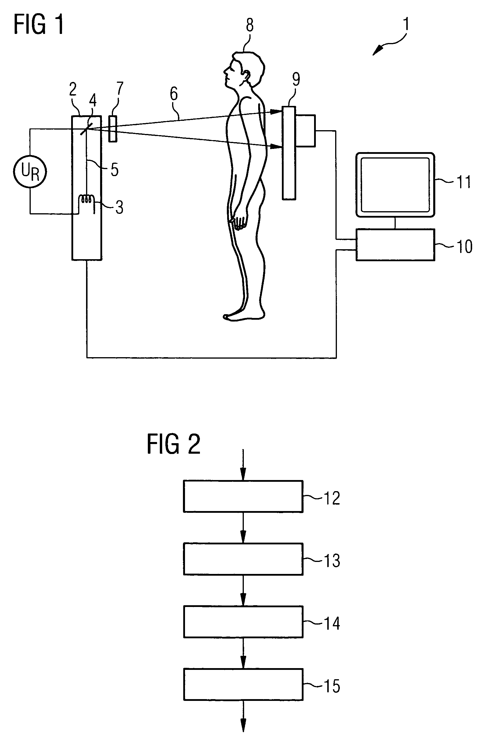
[0009]With said method, the optical thickness in a second energy range of the object being examined is approximately determined by the evaluation unit using a projection image recorded in a first high-energy range. The evaluation unit then sets recording parameters for recording the projection image in the second energy range as a function of the determined optical thickness of the object being examined. It is possible, using the determined optical thickness, to minimize the exposure to radiation of the object being examined and maximize the image quality of the projection image recorded in the second energy range, taking account of the required exposure time and the radiating power the radiation source is capable of producing. Overall, the exposure to radiation of the object being examined can thus be kept low.
[0010]In a preferred embodiment the optical thickness in the low-energy range of the object being examined is approximately determined by the evaluation unit using the projection image recorded in the high-energy range. The evaluation unit then sets the recording device's recording parameters, in particular those of the radiation source and detector, as a function of the determined optical thickness of the object being examined. Because the effective cross-section of the radiation quanta in terms of their impact on the material of the object being examined decreases as the quantum energy increases, a projection image recorded in the high-energy range will result in less exposure to radiation of the object being examined since fewer absorption processes take place than in the case of lower energy levels. Moreover, the recording parameters can only be poorly optimized in the case of high energy levels owing to the spectrum's wide distribution. The optical thickness in the low-energy range of the object being examined can, however, be approximately determined using the projection image recorded in the high-energy range. If the optical thickness in the low-energy range is known, recording parameters of the recording device that are matched to the optical thickness of the object being examined can be selected and set. The exposure to radiation tending to be higher during projection recording in the low-energy range can thereby be reduced to the extent necessary in keeping with the absorption characteristics of the object being examined.
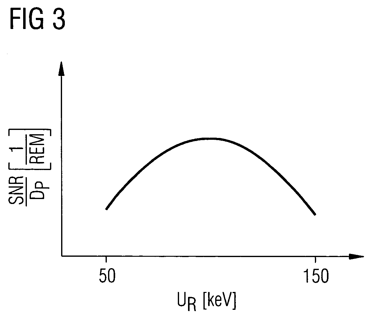
[0011]In a preferred embodiment the ratio between the signal-to-noise ratio of the projection image recorded in the low-energy range and the exposure to radiation of the object being examined is maximized through the choice of recording parameters in the low-energy range, taking account of the pre-specified exposure time and the radiating power the radiation source is capable of producing. A setting of said type will, taking account of the performance capability of the radiation source, enable the optimal image quality to be achieved with the exposure to radiation of the object being examined being minimized.
[0012]The recording parameters selected by the evaluation unit for recording the projection image in the low-energy range are read out by the evaluation unit from a predefined table preferably as a function of the determined optical thickness of the object being examined and required exposure time. The advantage thereof is that the recording parameters will not have to be recalculated every time, thus making the process of determining the recording parameters low in compute intensiveness. It is furthermore possible to incorporate experience-based knowledge into the tables.
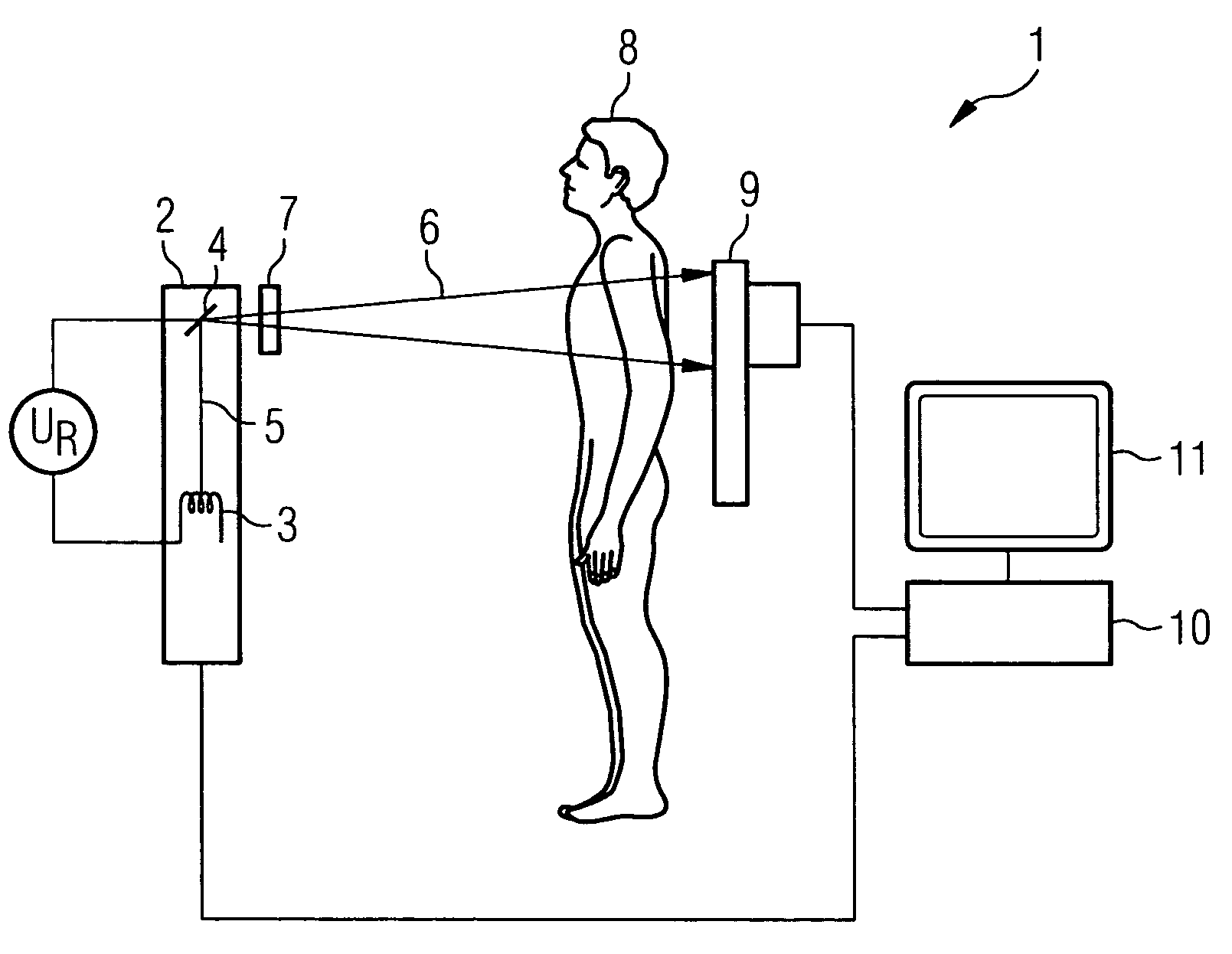
[0013]In a further preferred embodiment the radiation source is an x-ray source and the detector is an x-ray detector. If the x-ray source is an x-ray tube, then the recording parameters requiring to be set are the tube voltage, the tube current, the material and thickness of any preliminary filters that may be present, and the exposure time. Exposure to radiation in the case of medical applications can be not insubstantially reduced for a patient thanks to adaptive setting of the recording parameters for recording in the low-energy range. The image quality of the combined image can furthermore be increased.

Problems solved by technology

Moreover, the recording parameters can only be poorly optimized in the case of high energy levels owing to the spectrum's wide distribution.

Method used


Image

  • Method for recording projection image
  • Method for recording projection image
  • Method for recording projection image

Examples


Embodiment Construction

[0018]FIG. 1 shows an x-ray system 1 with which x-ray recordings for dual x-ray absorptiometry can be produced. The x-ray system 1 includes an x-ray tube 2 having a cathode 3 for emitting electrons. The cathode 3 is as a rule formed from an incandescent filament through which a heater current IR flows. The electrons emitted by the cathode 3 are accelerated toward an anode 4 with the aid of a tube voltage UR. That produces an electron beam 5 that impinges on the anode 4 in a focused spot. The electrons retarded in the anode 4 produce x-radiation 6 which first passes through a preliminary filter 7 to suppress the low-energy component. The preliminary filters 7 are as a rule copper plates that can be interposed having different thickness into the beam path of the x-radiation 6. The x-radiation 6 then penetrates a patient 8 being examined.

[0019]The x-radiation 6 that has passed through the patient 8 impinges on an x-ray detector 9 that produces an absorption image of the patient 8, with...


PUM


Abstract

In recording projection images for dual absorptiometry, the optical thickness of an object being examined is determined on the basis of a projection image recorded in a high-energy range, and parameters for recording a projection image in the low-energy range are selected as a function of the determined optical thickness. The ratio between image quality and the radiation dosage of the object being examined can be optimized thereby.

Description

CROSS REFERENCE TO RELATED APPLICATIONS[0001]This application claims priority of German application No. 10 2005 061 357.8 filed Dec. 21, 2005, which is incorporated by reference herein in its entirety.FIELD OF THE INVENTION[0002]The invention relates to a method for recording projection images of an object being examined, the projection images being recorded in different energy ranges and further processed to produce a combined image.BACKGROUND OF THE INVENTION[0003]A device of said type is known from U.S. Pat. No. 6,343,111 B1. Said known device includes a recording device having an x-ray source and an x-ray detector. Projection images of an object being examined can be recorded successively in a high-energy range and a low-energy range by means of said recording device. A combined image can then be produced by an evaluation unit from the projection images recorded in the high-energy and low-energy range.[0004]Because the absorption behavior of the irradiated material of the object...

Claims


Application Information

Patent Timeline
Patent Type & Authority Patents(United States)
IPC IPC(8): H05G1/64
CPCA61B6/4035A61B6/405A61B6/463A61B6/482A61B6/50
Inventor BERNHARDT, PHILIPP
Owner SIEMENS AG
Why Eureka
Try Eureka