Enhanced image processing method for the presentation of digitally-combined medical images

a technology of enhanced image processing and medical images, applied in the field of medical imaging, can solve problems such as strong distortions in the area, and achieve the effect of improving image processing

Active Publication Date: 2008-08-12
GE MEDICAL SYST GLOBAL TECH CO LLC
View PDF6 Cites 66 Cited by
  • Summary
  • Abstract
  • Description
  • Claims
  • Application Information

AI Technical Summary

Problems solved by technology

This presents a challenge because a wide range of anatomical thicknesses must be represented.
Conventional image-equalization algorithms may compensate for this effect, but extreme parameters must be used, leading to potentially strong distortions in the area of interest, as such algorithms are typically designed for the specific detector size.

Method used


Image

  • Enhanced image processing method for the presentation of digitally-combined medical images
  • Enhanced image processing method for the presentation of digitally-combined medical images
  • Enhanced image processing method for the presentation of digitally-combined medical images

Examples


Embodiment Construction

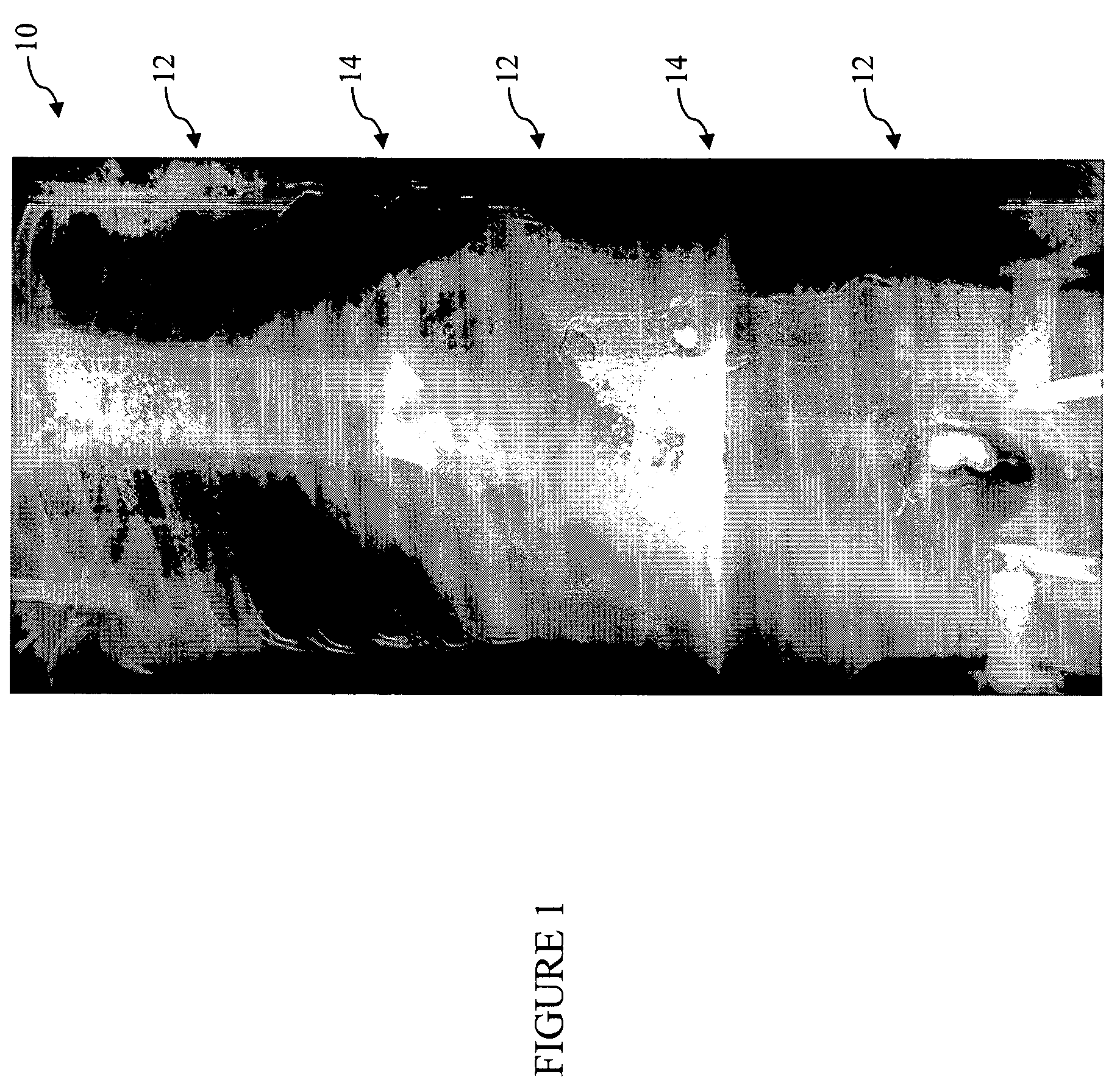
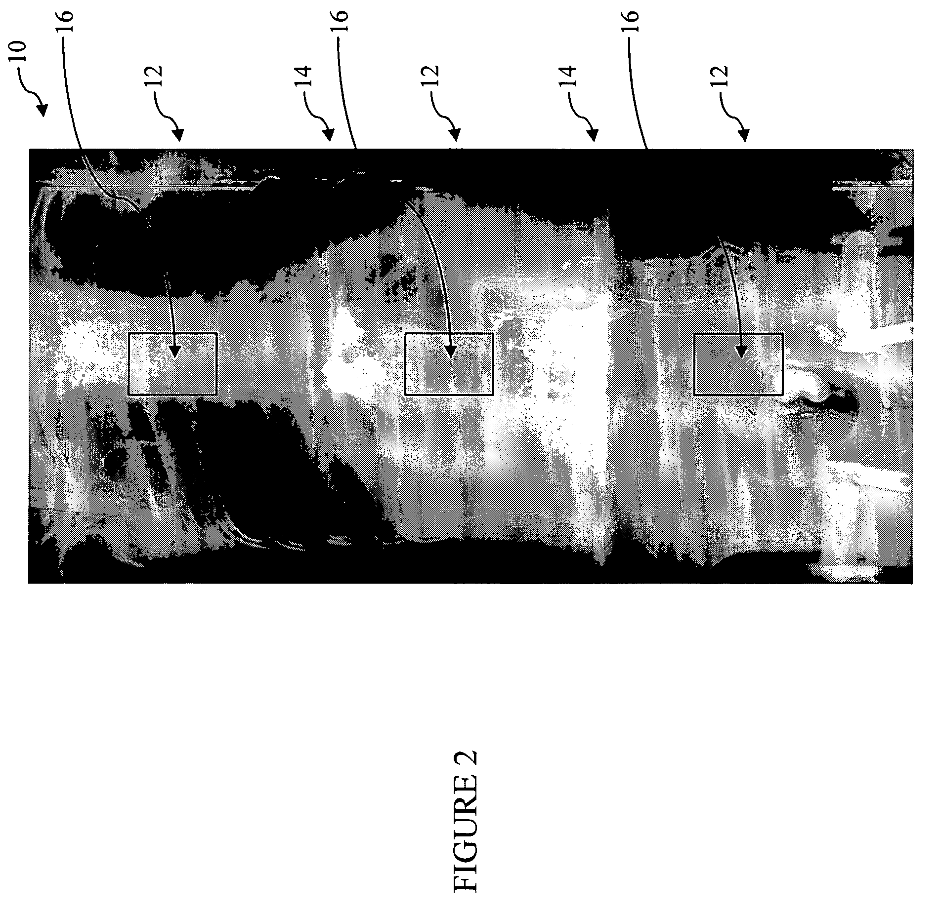
[0021]For the purposes of promoting an understanding of the present invention, reference will now be made to some preferred embodiments of the present invention, as illustrated in FIGS. 1-8, and specific language use to describe the same. The terminology used herein is for the purpose of description, and not limitation. The specific structural and functional details disclosed herein are not to be interpreted as limiting, but merely as a basis for the claims and for teaching one of ordinary skill in the art to variously employ the systems and methods of the present invention. Any modifications to or variations in the depicted systems and methods and such further applications of the principles of the present invention as would normally occur to one of ordinary skill in the art are considered to be within the spirit and scope of the present invention.

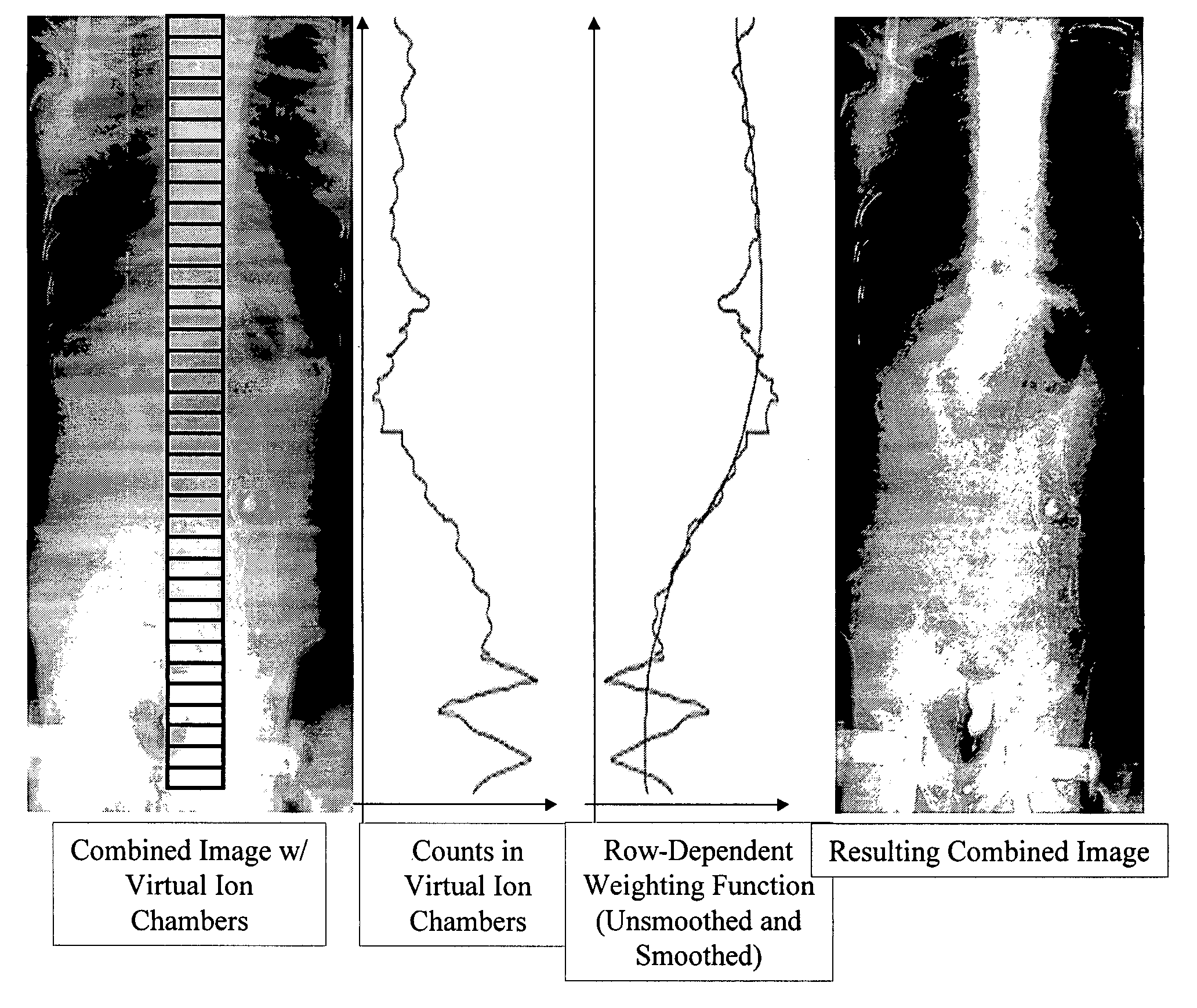
[0022]As described above, the flat-panel digital radiographic imaging detectors available today typically have a maximum imaging size of ...


PUM


Abstract

The present invention provides a method and associated system for processing a digital medical image. The method includes defining a plurality regions of an initial digital medical image, wherein the initial digital medical image is a combined initial digital medical image formed from the digital pasting of a plurality of individual initial digital medical images, and wherein the initial digital medical image is an exposure-normalized initial digital medical image; measuring an intensity for each of the plurality of regions of the initial digital medical image; deriving an intensity weighting function using the intensity measured for each of the plurality of regions of the initial digital medical image; and applying the intensity weighting function to the initial digital medical image to form a final digital medical image.

Description

FIELD OF THE INVENTION[0001]The present invention relates generally to the medical imaging field. More specifically, the present invention relates to an enhanced image processing method for the presentation of digitally-combined medical images.BACKGROUND OF THE INVENTION[0002]With applications ranging from diagnostic procedures to radiation therapy, the importance of high-performance medical imaging is immeasurable. As a result, new high-performance medical imaging technologies are continually being developed. Digital medical imaging technologies represent the future of medical imaging. Digital medical imaging systems are capable of producing far more accurate and detailed images of an anatomical object than conventional, film-based medical imaging systems. Such digital medical imaging systems also allow for image enhancement once an anatomical object has been scanned, further enhancing their usefulness.[0003]The flat-panel digital radiographic imaging detectors available today typi...

Claims


Application Information

Patent Timeline
Patent Type & Authority Patents(United States)
IPC IPC(8): G06K9/36G06K9/00G06K9/32
CPCG06T3/4038G06T5/009G06T2207/30004G06T2207/10116
Inventor BATTLE, VIANNEY PIERREUPPALURI, RENUKAWARP, RICHARD
Owner GE MEDICAL SYST GLOBAL TECH CO LLC
Why Eureka
Try Eureka