Method for recording and evaluating image data with the aid of a tomography machine

a tomography machine and image data technology, applied in tomography, material analysis using wave/particle radiation, instruments, etc., can solve the problems of increasing the administration of contrast medium, requiring a longer scanning time, and unable to use the attenuation value distribution of such x-ray images to deduce the material composition of an examination obj

Active Publication Date: 2008-11-25
SIEMENS HEALTHCARE GMBH
View PDF15 Cites 11 Cited by
  • Summary
  • Abstract
  • Description
  • Claims
  • Application Information

AI Technical Summary

Benefits of technology

[0015]An object of an embodiment of the present invention is to specify a method for recording and evaluating image data with the aid of a tomography machine which permits the image data to be recorded in conjunction with different spectral distributions for the purpose of evaluating the image data in accordance with the methods. Preferably, it avoids at least one of the above disadvantages.
[0018]By using a tomography machine having at least two recording systems, it is possible in accordance with the method of an embodiment to acquire spectral measured data in a single scan and use them to obtain, for example, quantitative additional information relating to the examination area, in particular to the materials contained therein, by way of the 2-spectra method or the ρ-Z projection. The problem of patient movement may be reduced or even eliminated, since the detectors assigned to the two X-ray tubes of the two recording systems acquire the measured or attenuation data at the same time. By comparison with the conventional methods for acquiring spectral measured data, the scanning time may be halved because, for example, the required spectral volumetric data are obtained in a single spiral scan. It is also possible to use a C arc system with at least two C arcs as a tomography machine. However, it is preferred to use an X-ray computer tomography machine having at least two recording systems, also denoted below as 2 tube detector CT system.
[0025]It is therefore possible to use this variant of the present method primarily to carry out advantageously angiographic CT examinations where it is necessary to segment bones in order to represent blood vessels near the bone. It is likewise desirable to suppress the image signal from soft part tissue when the contrast / noise ratio in the blood vessels filled with contrast medium is relatively low.
[0027]A further application of the last-named subtraction method is suggested in the case of CTAs for the purpose of representing the pulmonary perfusion, in the case of which CTAs there has hitherto likewise been a need for an additional native scan to remove the soft part background. The consequence of this is that the movement of the blood vessels in the lung can substantially impair the quality of the subtraction CTA. Because the detectors assigned to the two X-ray tubes acquire the attenuation data simultaneously in the present method, the spectral data are measured simultaneously, thereby avoiding problems due to a movement of the blood vessels.

Problems solved by technology

The attenuation value distribution of such X-ray images cannot, however, be used to deduce the material composition of an examination object, since the X-ray absorption is determined both by the effective atomic number of the material and by the material density.
However, these techniques favor the disturbing influence of patient movement, require a longer scanning time and also an increased administration of contrast medium in the case of CT examinations based on contrast medium.

Method used


Image

  • Method for recording and evaluating image data with the aid of a tomography machine
  • Method for recording and evaluating image data with the aid of a tomography machine
  • Method for recording and evaluating image data with the aid of a tomography machine

Examples


Embodiment Construction

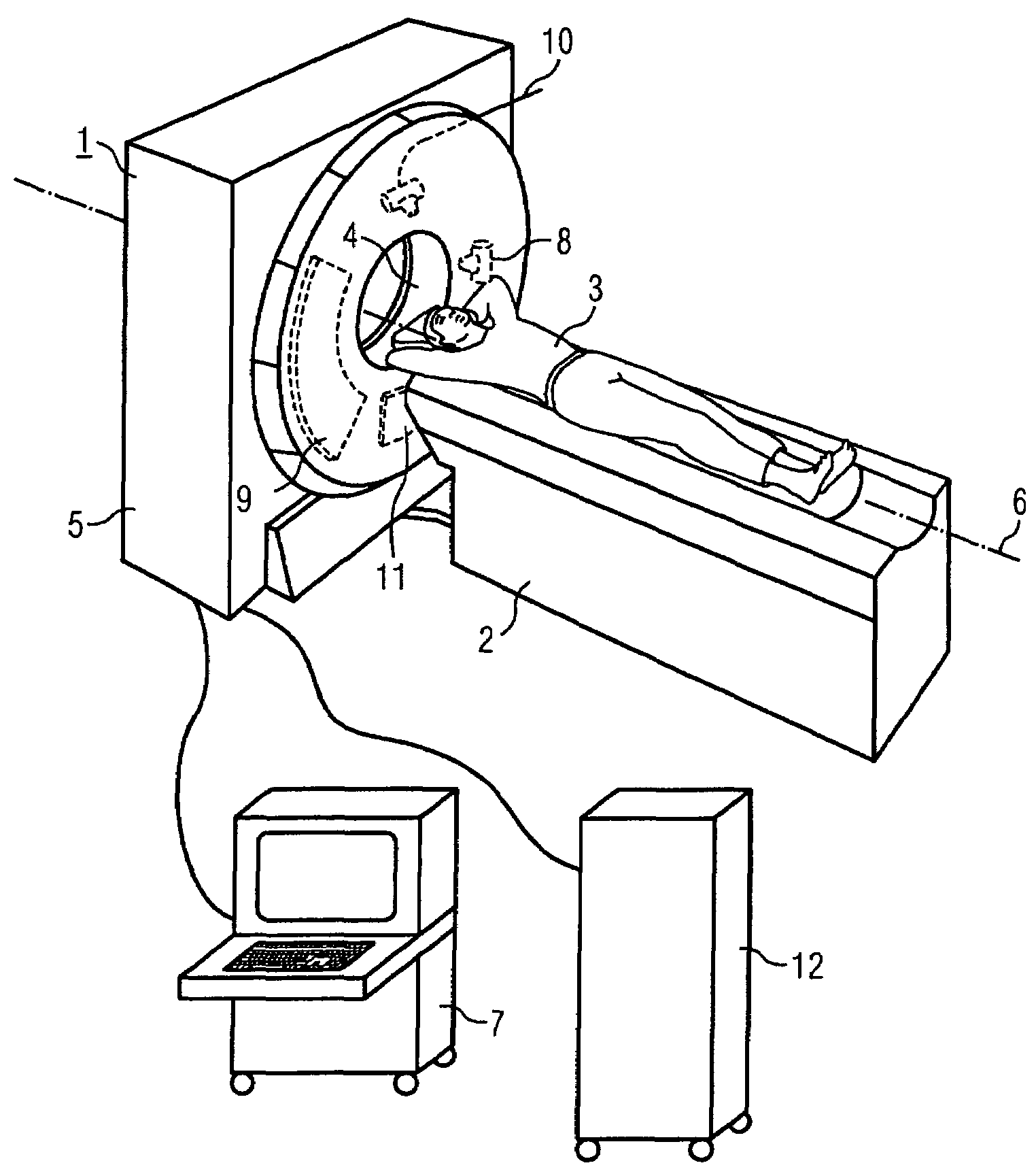
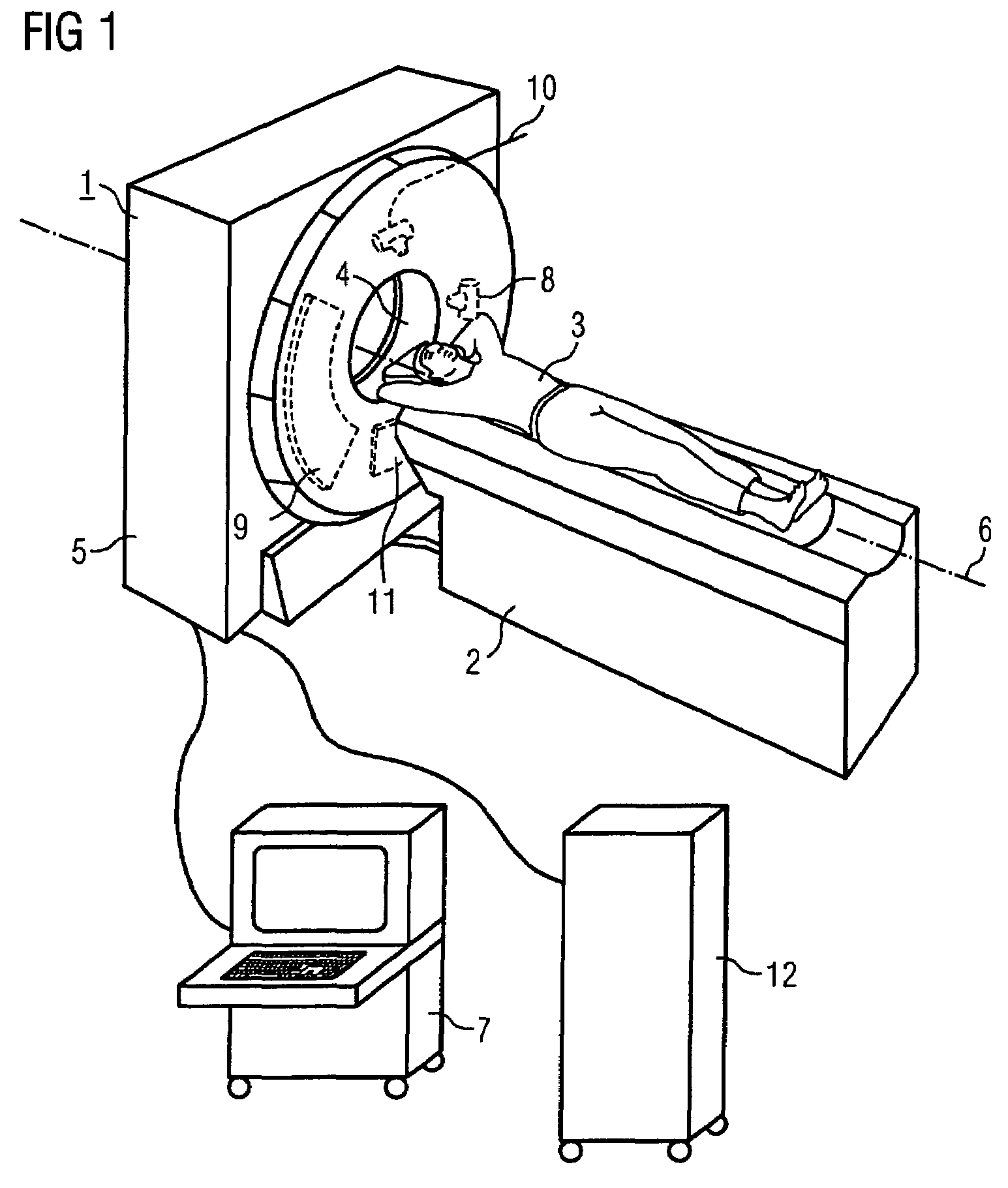
[0035]FIG. 1 shows a tomography machine 1, in the present example an X-ray computed tomography machine, having an assigned support device 2 for receiving and supporting a patient 3. The patient 3 with the desired examination area can be introduced into an opening 4 in the housing 5 of the tomography machine 1 by way of a moveable table plate of the supporting device 2. During a spiral scan, the supporting device 2 is useful, however, to effect continuous axial feeding. A gantry (not visible in FIG. 1) can be rotated in the interior of the housing 5 at high speed about a rotation axis 6 running through the patient 3. An operating unit 7 is also present for operating the tomography machine 1.

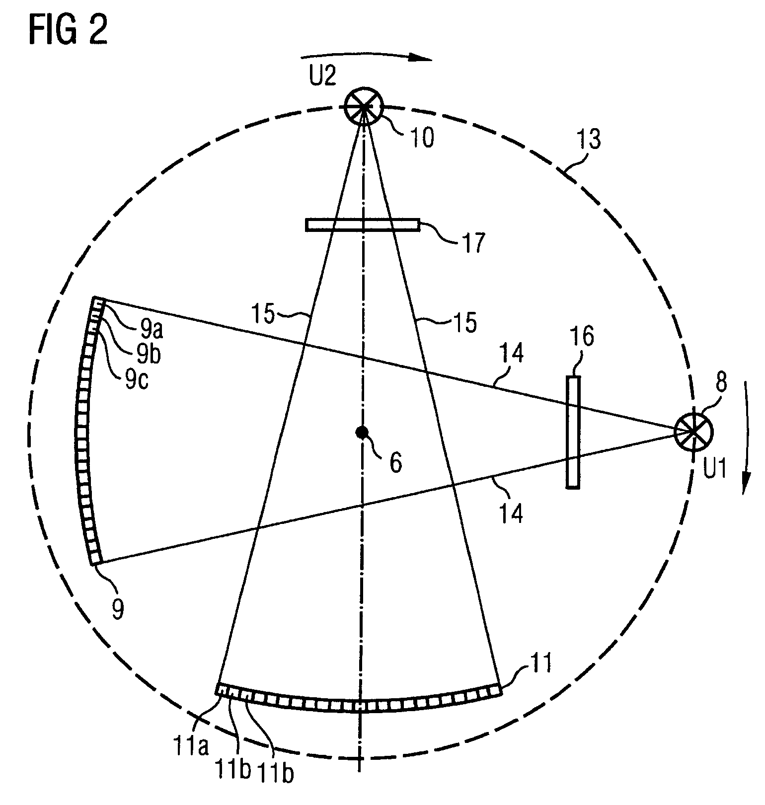
[0036]The present tomography machine 1 has two recording systems on the gantry, and these respectively include an X-ray tube 8 or 10 respectively, and a multirow X-ray detector 9 and 11 respectively. The arrangement of the two X-ray tubes 8, 10 and the two detectors 9, 11 on the gantry is fixed du...


PUM

PropertyMeasurementUnit
angleaaaaaaaaaa
atomic numberaaaaaaaaaa
tube voltageaaaaaaaaaa

Abstract

A method is for recording and evaluating image data with the aid of a tomography machine. At least two recordings with different spectral distribution are made of an examination area of an object. Further, measured data obtained from the two recordings are evaluated such that additional information relating to the examination area and / or a specific representation of the image of the examination area are / is obtained from the different spectral distributions. The tomography machine includes at least two separate recording systems. Further, it is operated such that the two recording systems operate with a different spectral distribution. As such, the additional information and the specific representation of an image can be obtained in conjunction with a reduced scanning time.

Description

[0001]The present application hereby claims priority under 35 U.S.C. §119 on German patent application numbers DE 10 2004 004 295.0 filed Jan. 28, 2004, the entire contents of which is hereby incorporated herein by reference.FIELD OF THE INVENTION[0002]The present invention generally relates to a method for recording and evaluating image data with the aid of a tomography machine. Preferably, at least two recordings with different spectral distribution are made of an examination area of an object. Measured data obtained from the two recordings are then evaluated such that additional information relating to the examination area and / or a specific representation of the image of the examination area are / is obtained from the different spectral distributions.BACKGROUND OF THE INVENTION[0003]The result of radiographic methods such as, for example, computed tomography, mammography, angiography, the X-ray inspection technique or comparable methods, is firstly the representation of the attenua...

Claims


Application Information

Patent Timeline
Patent Type & Authority Patents(United States)
IPC IPC(8): G06K9/00G01N23/00A61B6/03G01N23/06G06T1/00G21K1/12H05G1/60
CPCA61B6/032A61B6/4035A61B6/405A61B6/482Y10S378/901
Inventor BRUDER, HERBERTFLOHR, THOMASHEISMANN, BJOERNRAUPACH, RAINERSTIERSTORFER, KARL
Owner SIEMENS HEALTHCARE GMBH
Why Eureka
Try Eureka