X-ray computed tomography apparatus

a computed tomography and computed tomography technology, applied in tomography, instruments, diagnostic recording/measuring, etc., can solve the problems of unstable actual cardiac cycle of a subject (patient), inability to determine an x-ray application period, and inability to change the cardiac cycle corresponding to several heartbeats. , to achieve the effect of preventing the omission of a part of the projection data

Active Publication Date: 2010-05-11
TOSHIBA MEDICAL SYST CORP
View PDF16 Cites 15 Cited by
  • Summary
  • Abstract
  • Description
  • Claims
  • Application Information

AI Technical Summary

Benefits of technology

[0011]It is an object of the present invention to prevent the omission of part of projection data required for image reconstruction in an X-ray computed tomography apparatus which can perform ECG gated reconstruction.

Problems solved by technology

However, the actual cardiac cycle of a subject (patient) is unstable, and a cardiac cycle corresponding to several heartbeats is not constant even before scanning.
It is therefore not practical to determine an X-ray application period by using only a cardiac cycle corresponding to one heartbeat.
In examination of a fast-moving part using an X-ray computed tomography apparatus, and in particular, cardiac examination, one of the important challenges is to improve the time resolution of images.
However, arrhythmia or the like sometimes causes a situation in which projection data necessary for image reconstruction in a desired cardiac time phase has not been acquired, or projection data has not been acquired with a high S / N ratio.
In such a situation, the image quality of a reconstructed image deteriorates.
Furthermore, there is no means to check the above situation before reconstruction.

Method used


Image

  • X-ray computed tomography apparatus
  • X-ray computed tomography apparatus
  • X-ray computed tomography apparatus

Examples


first embodiment

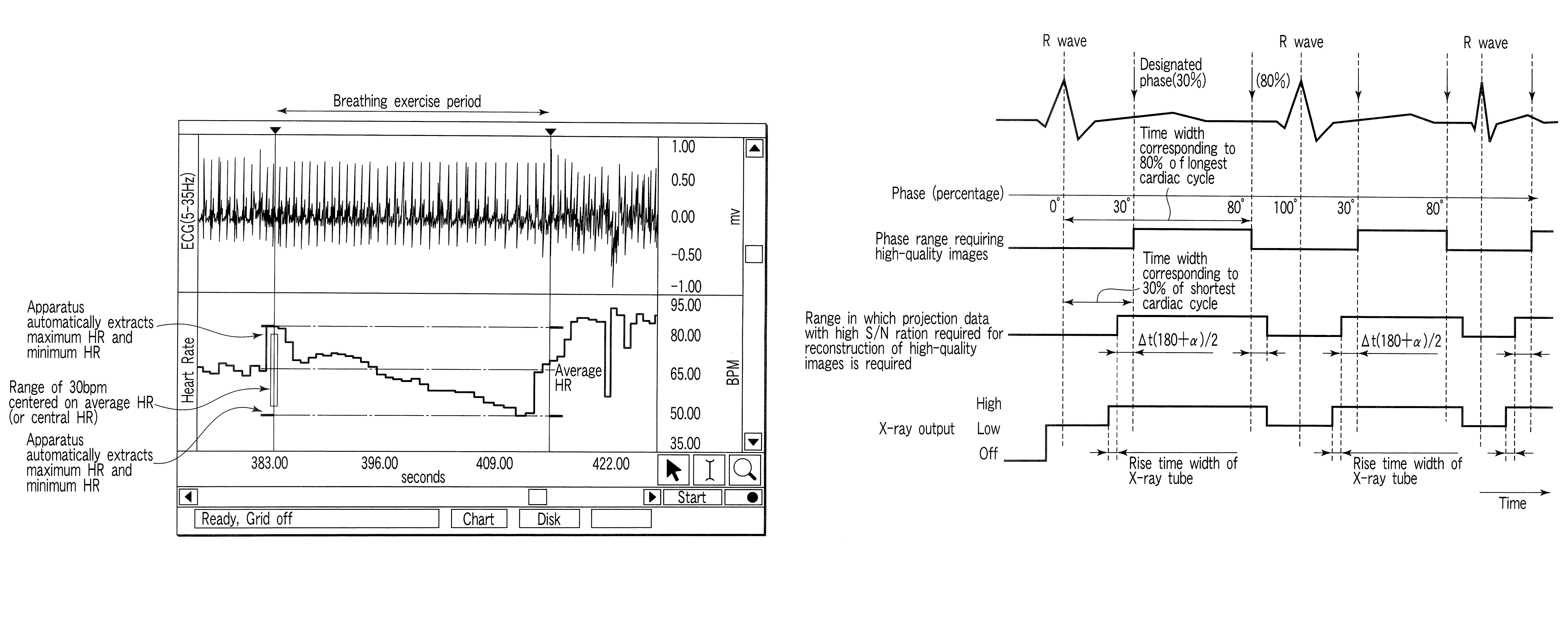
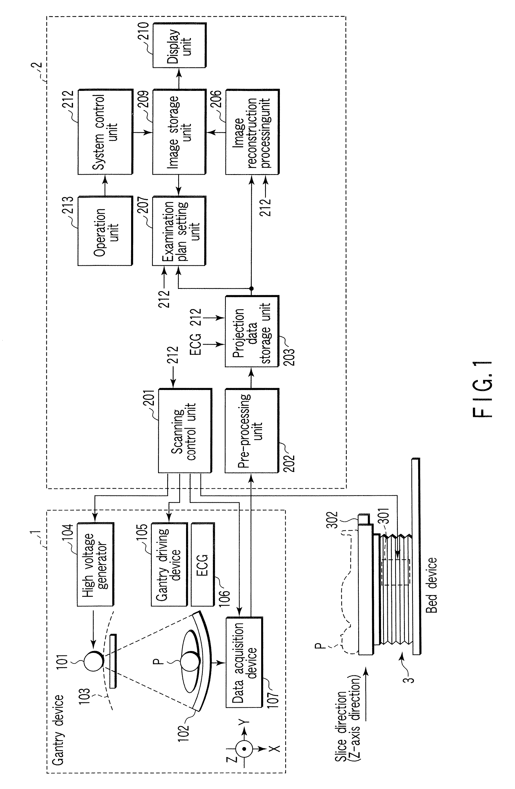
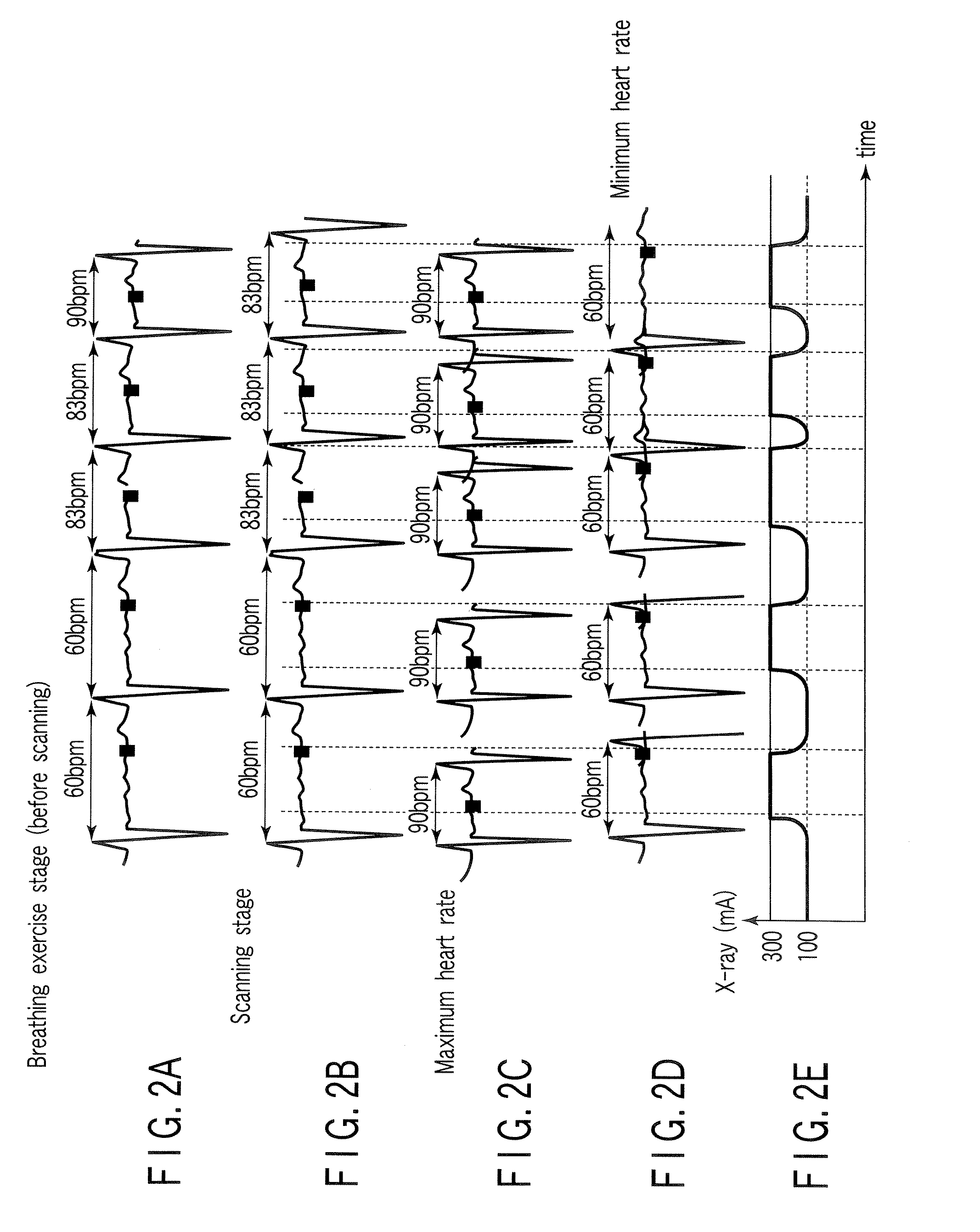
[0038]The first embodiment of an X-ray computed tomography apparatus according to the present invention will be described below with reference to the views of the accompanying drawing. Note that X-ray computed tomography apparatuses include various types of apparatuses, e.g., a rotate / rotate-type apparatus in which an X-ray tube and X-ray detector rotate together around a subject to be examined, and a stationary / rotate-type apparatus in which many detection elements are arrayed in the form of a ring or plane, and only an X-ray tube rotates around a subject to be examined. The present invention can be applied to either type. In this case, the rotate / rotate type, which is currently the mainstream, will be exemplified. In order to reconstruct one-slice tomogram data, projection data corresponding to one rotation around a subject to be examined, i.e., about 360°, is required (full reconstruction method), or projection data corresponding to 180°+α (α: fan angle) is required in the half r...

second embodiment

[0136]The second embodiment of an X-ray computed tomography apparatus (also called as an X-ray CT or CT scanner) according to the present invention will be described below with reference to the views of the accompanying drawing. Note that X-ray computed tomography apparatuses include various types of apparatuses, e.g., a rotate / rotate-type apparatus in which an X-ray tube and X-ray detector rotate together around a subject to be examined, and a stationary / rotate-type apparatus in which many detection elements are arrayed in the form of a ring, and only an X-ray tube rotates around a subject to be examined. The present invention can be applied to either type. In this case, the rotate / rotate type, which is currently the mainstream, will be exemplified.

[0137]In addition, reconstruction methods include the full reconstruction method, half reconstruction method, and segment reconstruction method. These reconstruction methods differ in angle range required to reconstruct one-slice tomogra...


PUM

PropertyMeasurementUnit
timeaaaaaaaaaa
fan angleaaaaaaaaaa
tube currentaaaaaaaaaa

Abstract

An X-ray computed tomography apparatus includes an X-ray source, a high voltage generating unit, an X-ray detector detecting X-rays transmitted through a subject to generate projection data, a storage unit storing the projection data in association with electrocardiographic data of the subject, a setting unit setting a specific cardiac phase in accordance with an operator's instruction, a reconstruction unit reconstructing a tomogram based on projection data sets acquired in specific periods centered on the specific cardiac phase throughout cardiac cycles, a period extending unit extending the specific period on the basis of a heart rate fluctuation range of the subject, and a control unit controlling the high voltage generating unit to generate a relatively high dose of X-rays in the extended specific period and generate a relatively low dose of X-rays in a period other than the extended specific period.

Description

CROSS-REFERENCE TO RELATED APPLICATIONS[0001]This application is based upon and claims the benefit of priority from prior Japanese Patent Applications No. 2005-259614, filed Sep. 7, 2005; and No. 2005-285863, filed Sep. 30, 2005, the entire contents of both of which are incorporated herein by reference.BACKGROUND OF THE INVENTION[0002]1. Field of the Invention[0003]The present invention relates to an X-ray computed tomography apparatus which can perform ECG gated reconstruction.[0004]2. Description of the Related Art[0005]An X-ray computed tomography apparatus provides information about a subject to be examined in the form of images on the basis of the intensities of X-rays transmitted through the subject, and serves as an important role in many medical activities such as disease diagnosis, medical treatment, and surgical operation planning.[0006]In examination of the heart which moves fast, ECG gated scanning is performed. ECG gated scanning is performed to acquire ECG gated signal...

Claims


Application Information

Patent Timeline
Patent Type & Authority Patents(United States)
IPC IPC(8): H05G1/62G01N23/083
CPCA61B6/032A61B6/541A61B6/542A61B6/503A61B5/0245A61B6/027A61B6/405
Inventor NAGATA, KIYOSHITSUYUKI, MASAHARU
Owner TOSHIBA MEDICAL SYST CORP
Why Eureka
Try Eureka