Method and apparatus for extracting cerebral ventricular system from images

a cerebral ventricular system and image technology, applied in image enhancement, image analysis, instruments, etc., can solve the problems of inability to avoid inclusion of non-ventricular csp regions, inability to work, and dependence of this algorithm on coarse binary brain segmentation image and coarse white matter segmentation image, etc., to achieve more robust and flexible effects

Inactive Publication Date: 2010-07-13
AGENCY FOR SCI TECH & RES
View PDF11 Cites 8 Cited by
  • Summary
  • Abstract
  • Description
  • Claims
  • Application Information

AI Technical Summary

Problems solved by technology

In reality, however, this does not work and many factors should be taken into account such as noise, spatial and contrast resolutions of the scan.
In particular, the method has to handle a partial volume effect causing “leakages” from the ventricles to the extraventricular space, and intraventricular and extraventricular pathology distorting the normal anatomy of the ventricular system.
One of the limitations of this algorithm is its dependence on the coarse binary brain segmentation image and the coarse white matter segmentation image.
Another limitation is that it is not able to avoid inclusion of non-ventricular CSP regions although some anatomical knowledge of the ventricular system has been incorporated into the method to guide the location of the seed point(s) and searching subregions.
Furthermore, the algorithm described is not able to extract the complete ventricular system as the fourth ventricle is not extracted.
Several artificial boundaries are required when extracting the third ventricle resulting in some amount of manual editing needed to obtain precise results.
In addition, the algorithm is time consuming to run (about 5 to 20 min on a Pentium3 450 MHz PC for a brain volume).
It needs manual editing of the resulting outlines due to irregularities in automatically generated outlines caused by the partial volume effect and low contrast.
As such, the algorithm is dependent on the digital atlas and is time consuming to run (5-10 minutes).
The algorithm works best with images of normal subjects and is time consuming to run.
The major limitations of this method are that it can only extract parts of ventricles and that it is time consuming.
There are several problems with this approach.
The technique is sensitive to the initial position of the atlas, that is, if the initial rigid alignment is off by too much, the elastic match may perform poorly.
The presence of neighbouring features may also cause matching problems, for example, the atlas may warp to an incorrect boundary.
Without user interaction, the atlas can have difficulty converging to complicated object boundaries.
Although the statistical point model is able to capture considerable variability for the lateral ventricular boundary, it is not easy to build the statistical model specific to all classes of the ventricular system.
Furthermore, this approach is time consuming.
From the results presented, the algorithm does not appear to be able to extract the ventricular system accurately, or to maintain the connectivity automatically.
One limitation is that it can only handle brain with no gross anatomic abnormality.
In addition, the manual identification of contours of neuroanatomical structures is tedious, time-consuming and vulnerable to inter-personal variations.
However, if the patient's brain has been deformed too much, for example, due to high variability or disease, the model does not work well.
In addition to the human interaction for initial information, the algorithm is not able to maintain the connectivity automatically.
One limitation of the algorithm is that it is restricted to slice images where the brain parenchyma and CSF spaces form connected regions.
In addition, it is difficult to locate, automatically, the threshold pixel intensity from the intersection of Gaussian fitting curves because the number of modals of the histogram of a ROI is difficult to determine.
However, this method it is too crude for ventricular volume analysis.
Despite the numerous approaches proposed to solve the problem of extracting the human cerebral ventricular system, the above-described methods suffer from disadvantages and, as such, are not suitable for clinical use.
This is due to the necessity for human intervention, the inability of these methods to handle pathological and abnormal cases, and / or the inability to extract the complete ventricular system.
Moreover, the existing methods are too slow to be accepted clinically.

Method used


Image

  • Method and apparatus for extracting cerebral ventricular system from images
  • Method and apparatus for extracting cerebral ventricular system from images
  • Method and apparatus for extracting cerebral ventricular system from images

Examples


Embodiment Construction

1. Abbreviations Used in the Disclosure

[0051]The following abbreviations are used in the present specification:[0052]AC—anterior commissure[0053]PC—posterior commissure[0054]MSP—midsagittal plane[0055]VL—lateral ventricle[0056]V3—third ventricle[0057]V4—fourth ventricle[0058]VLL—left lateral ventricle[0059]VLR—right lateral ventricle[0060]VLL-B—body of the left lateral ventricle (including the anterior and posterior horns)[0061]VLR-B—body of the right lateral ventricle (including the anterior and posterior horns)[0062]VLL-I—inferior (temporal) horn of the left lateral ventricle[0063]VLR-I—inferior (temporal) horn of the right lateral ventricle[0064]AC-PC—intercommissural (axial) plane passing through the AC and PC[0065]VAC—coronal plane passing through the AC[0066]VPC—coronal plane passing through the PC[0067]CSF—cerebrospinal fluid[0068]GM—grey matter[0069]WM—white matter[0070]ROI—two-dimensional region of interest[0071]2D—two-dimensional (or two dimensions)[0072]3D—three-dimension...


PUM


Abstract

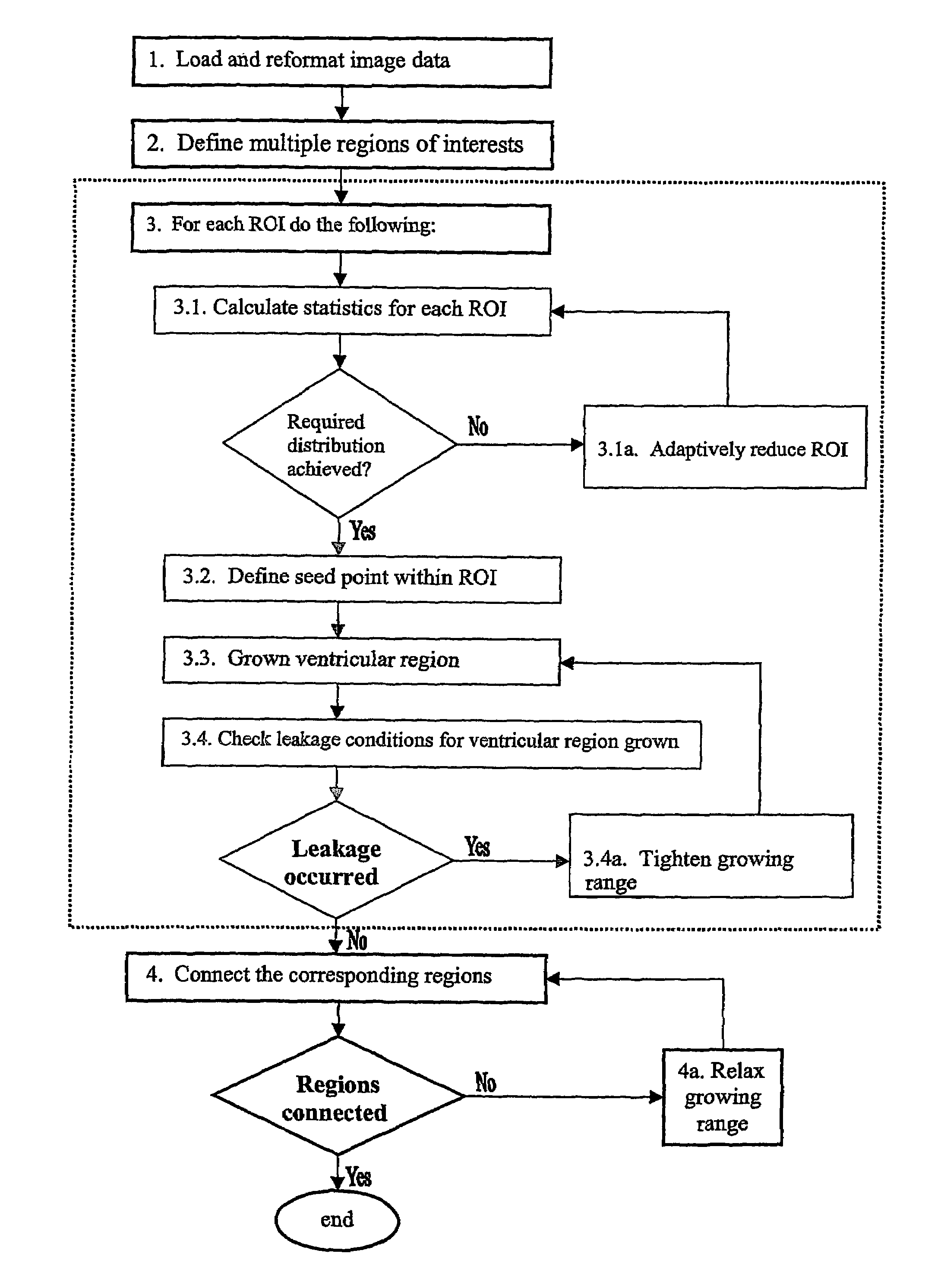
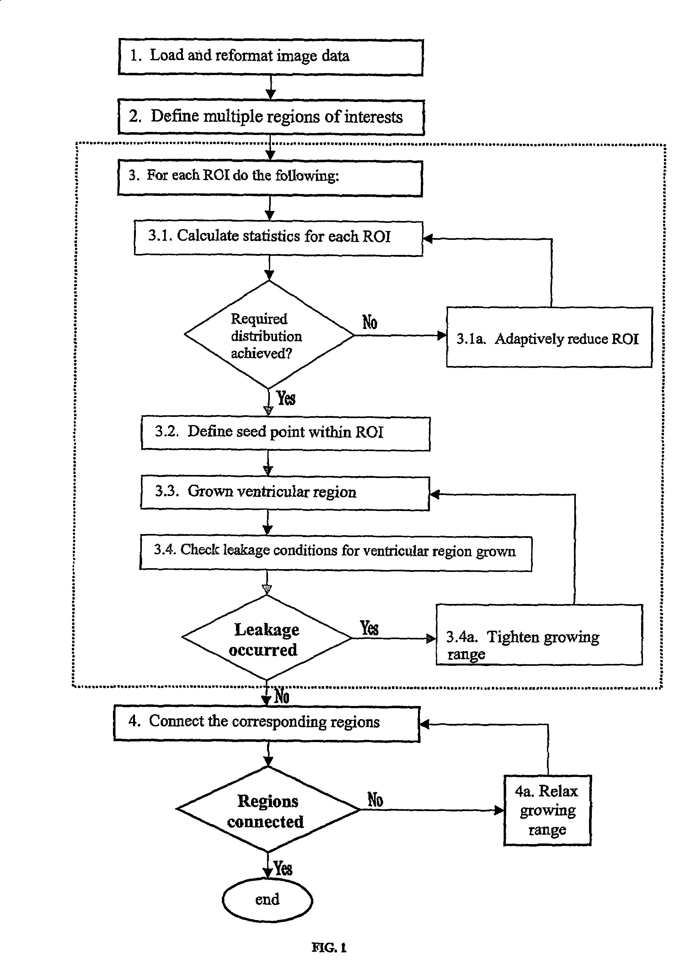
A method and apparatus for extracting a cerebral ventricular system from images of one or more cerebral ventricular regions comprises defining multiple regions of interest (ROI) in the images, defining seed points within each ROI, growing images of ventricular regions while correcting for leakages into extraventricular space and connecting the ventricular regions grown.

Description

FIELD OF INVENTION[0001]The present invention relates to a method and appara-tus for extracting cerebral ventricular system information from images of one or more cerebral ventricular regions. In particular, the present invention relates to extracting the human cerebral ventricular system from radiological images.BACKGROUND OF THE INVENTION[0002]The human cerebral ventricular system contains four intercommunicating chambers, the left lateral ventricle, the right lateral ventricle, the third ventricle, and the fourth ventricle. The ventricles contain cerebrospinal fluid (CSF) and changes in CSF volume and shape are associated with several neurological diseases such as congenital anomalies, post-traumatic disorders, pseudo-tumors, neuro-degenerative diseases, inflammatory diseases, headache, cognitive dysfunction / psychiatric diseases and post-operative changes. Quantification of the degree of dilatation of ventricles is important in the diagnosis of various diseases and for measuring ...

Claims


Application Information

Patent Timeline
Patent Type & Authority Patents(United States)
IPC IPC(8): G06K9/00G06T5/00G06T7/60G06V10/25
CPCG06K9/3233G06K9/342G06T7/0081G06K2209/05G06T2207/10088G06T2207/20141G06T2207/20156G06T2207/30016G06T7/11G06T7/187G06V10/25G06V10/267G06V2201/03
Inventor NOWINSKI, WIESLAW LUCJANXIA, YANAZIZ, AAMERHU, QINGMAO
Owner AGENCY FOR SCI TECH & RES
Why Eureka
Try Eureka