Methods and devices for the non-thermal, electrically-induced closure of blood vessels

a non-thermal, electrically induced technology, applied in the field of methods and devices for the non-thermal, electrically induced closure of blood vessels, can solve the problems of inability to achieve the effect limited effectiveness of many of these agents, and inability to achieve the effect of minimal electrochemical damag

Active Publication Date: 2012-01-31
THE BOARD OF TRUSTEES OF THE LELAND STANFORD JUNIOR UNIV
View PDF21 Cites 144 Cited by
  • Summary
  • Abstract
  • Description
  • Claims
  • Application Information

AI Technical Summary

Benefits of technology

[0014]One object of the invention is to selectively induce hemostasis within blood vessels by creating thrombosis within the vessels with minimal side effects, fewer steps and less discomfort to the patient than has heretofore been possible.

Problems solved by technology

It has been shown that a reduction in tumor blood flow reduces nutrients to the tumor and causes accumulation of catabolite products and extracellular acidification, all of which result in a cascade of tumor cell death.
However, the effectiveness of many of these agents is limited by the risk of unacceptable system toxicity (G. Sersa, M. Cemasar, C. S. Parkins and D. J. Chaplin: Tumor blood flow changes induced by application of electric pulses, European Journal of Cancer 35, N.
However, complete and permanent hemostasis has not yet been achieved by these methodologies.
Mechanical clamping of the tumor-supporting vasculature has also been proposed (Denekamp J, Hill S. A., Hobson B., Vascular occlusion and tumor cell death, Eur. J. Cancer Clin. Oncol. 1983, 19, 271-275), however, such technique may be impractical due to the extremely twisted and branched nature of tumor vasculature.
While not often of physical concern, they can be unsightly and cause emotional distress to the patient.
The injected substance injures the interior walls of the capillary causing it to shrink or disappear.
Unfortunately, this treatment can be painful, only partially effective, and usually requires about one to two months before improvement can be seen.
In addition, undesirable side effects can occur, such as echymotic or hyperpigmented marks, which may take months to completely fade away.
Where the affected skin area is relatively deep, the more superficial capillaries absorb the majority of the light energy and the remaining energy is insufficient to effectively treat the deeper vessels (referred to as “shadowing”).
This problem can be solved to some degree by use of less absorbent wavelengths, however, this is at the sake of a reduced ability to localize heat, which may necessitate longer treatments and / or multiple treatments which are both expensive and time-consuming.
Additionally, laser therapy does not work as well with patients having a darker skin pigment as the epidermal melanin absorbs a significant portion of the light to which it is exposed, thus, reducing the amount of light that is able to reach the blood.
The increase in the intensity of the laser required to compensate for interference from tissue and melanin may lead to thermal injury of the skin and to post-inflammatory pigment changes.
However, current electrosurgical devices and procedures are based on the thermal denaturation and coagulation of tissues and still suffer from significant thermal damage to surrounding tissue, and an inability to accurately control the depth of necrosis in the tissue being treated.
Additionally, the application of current to tissue results in electrochemical reactions which lead to the accumulation of toxic products on the electrodes that may cause cytolysis of the surrounding tissue (Peterson H. I., Tumor Blood Circulation Angiogenesis, Vascular Morphology and Blood Flow of Experimental and human tumors, Florida, CRC Press, 1979, 1-229).
In addition, hydrolysis on the electrodes emits gases which may interfere with current transmission, making the treatment unpredictable and unstable (S. Guarini, A Highly Reproducible Model of Arterial Thrombosis in Rats, Journal of Pharmacological and Toxicological Methods, 35 (1996) pp 101-105).

Method used


Image

  • Methods and devices for the non-thermal, electrically-induced closure of blood vessels
  • Methods and devices for the non-thermal, electrically-induced closure of blood vessels
  • Methods and devices for the non-thermal, electrically-induced closure of blood vessels

Examples


example

[0045]The following example is put forth so as to provide those of ordinary skill in the art with an exemplary disclosure and description of how to employ the present invention, and is not intended to limit the scope of what the inventor regards as his invention nor is it intended to represent that the experiments below are all or the only experiments performed. Efforts have been made to ensure the accuracy of the data, however, some experimental errors and deviations should be accounted for.

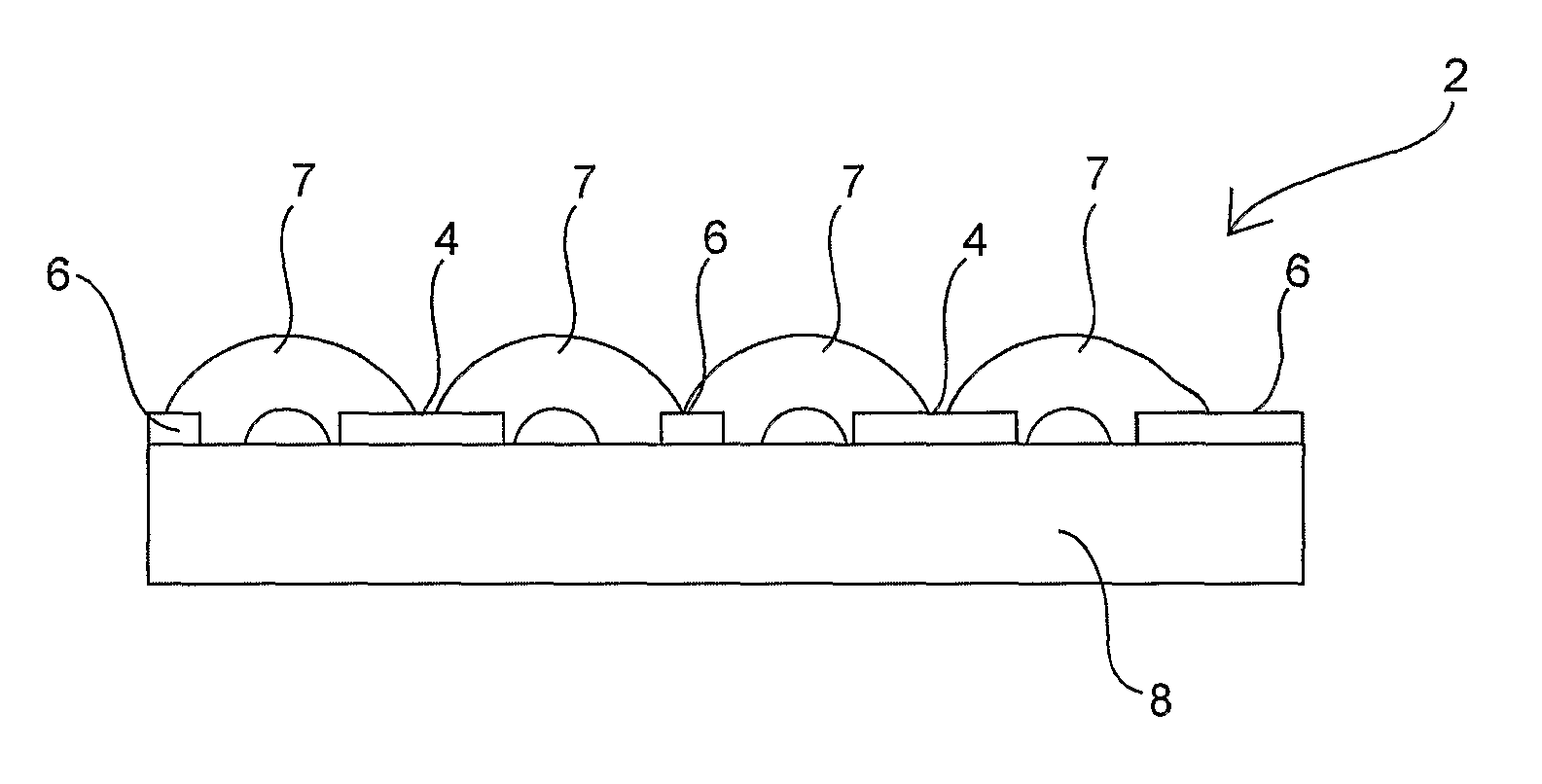
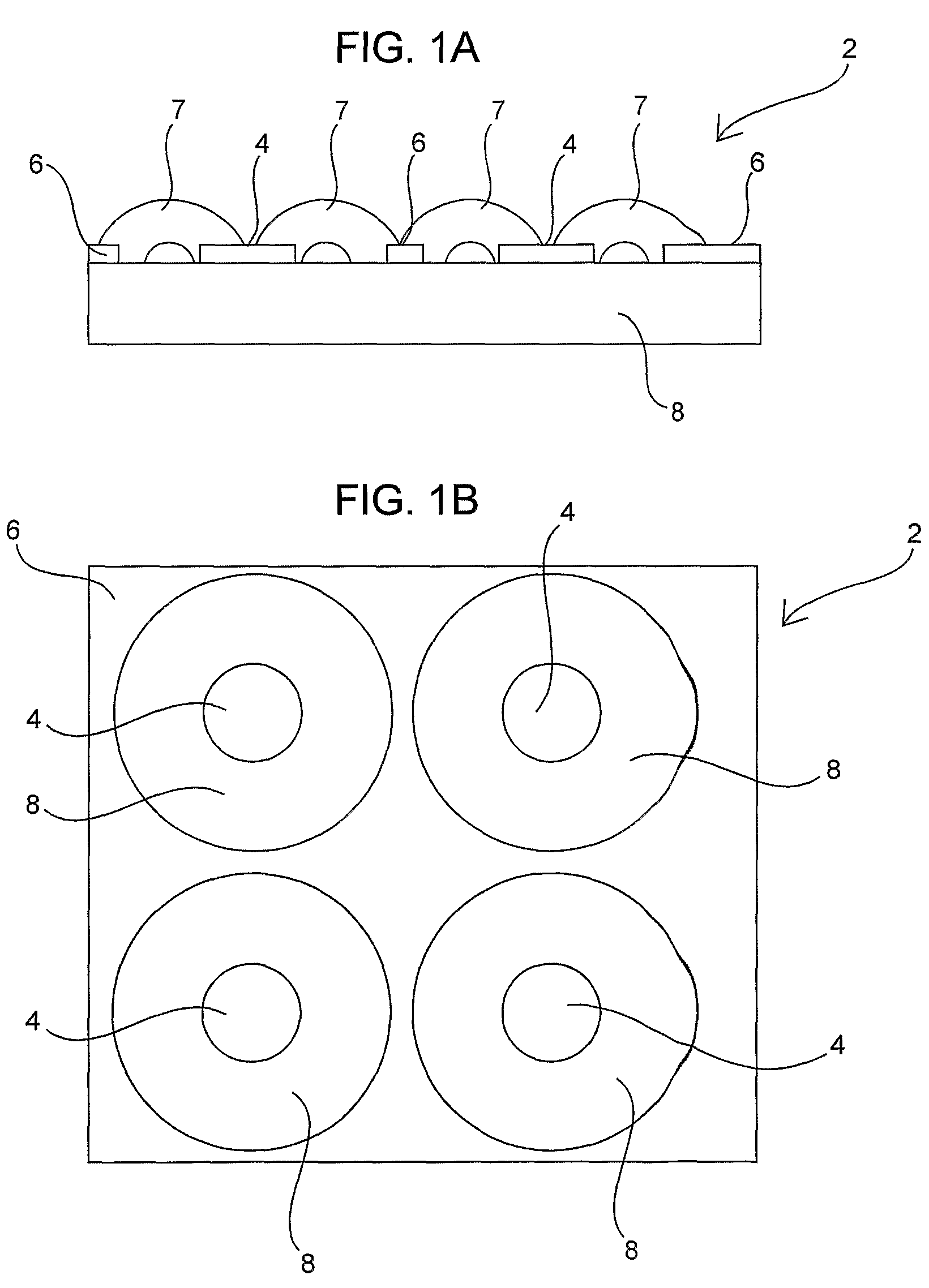
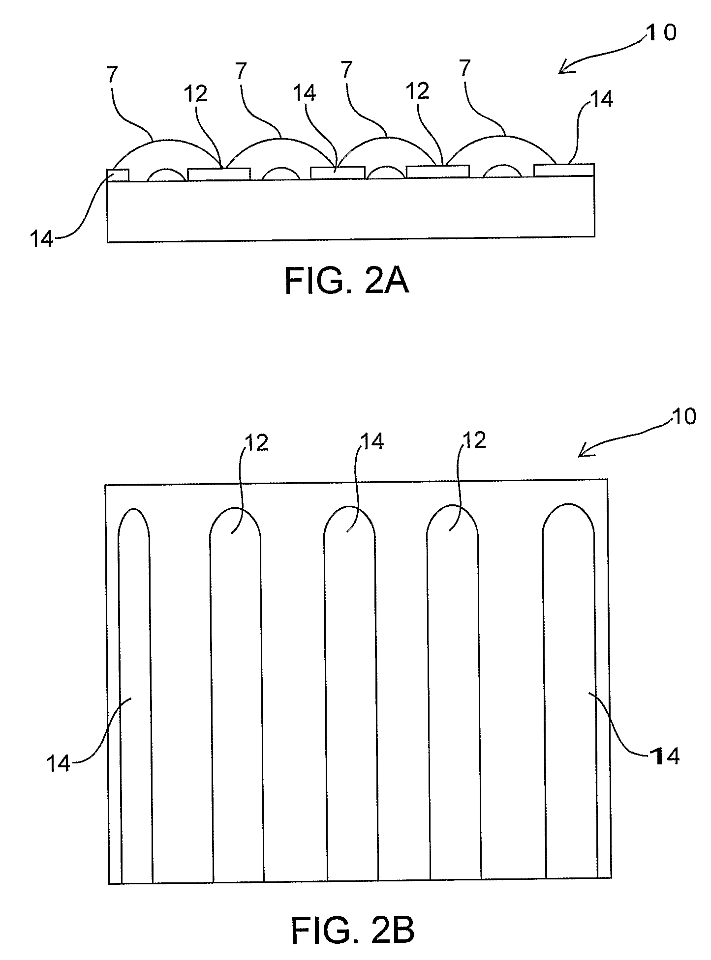
[0046]A chorioallantoic membrane (CAM) of a chicken embryo 17 days into the incubation cycle was used for performing the experiments. Various blood vessels (three arteries and three veins) of the CAM were selected for treatment, where the vessels were of varying diameters. An electrode of 2 mm in length, 300 μm in width and 50 μm in thickness was used. Using the electrode assembly, a selected minimum threshold voltage of 100 V was applied in biphasic (having positive and negative phases in the p...


PUM


Abstract

Methods and devices for the non-thermal, electrically-induced temporary or permanent closure of blood vessels. The subject methods and devices employ pulsed electrical energy according to a defined regime to effect controlled occlusion of targeted blood vessels without heating the vessel and with minimal damage to adjacent tissue. The extent of vessel closure, i.e., temporary (vasoconstriction) or permanent (thrombosis), is controlled based on the manipulation of various parameters of the electrical stimulation regime as well as the configuration of the electrodes used to apply the regime.

Description

FEDERALLY-SPONSORED RESEARCH OR DEVELOPMENT[0001]This invention was made with Government support under contract EY012888awarded by the National Institutes of Health. The Government has certain rights in this invention.CROSS-REFERENCE TO RELATED APPLICATIONS[0002]This application is a National Phase filing under 35 U.S.C. §371 of International Application No. PCT / US2005 / 033856 filed Sept. 20, 2005, which claims priority to U.S. Provisional Patent Application No. 60 / 612,835 filed Sep. 24, 2004, the contents of which are incorporated herein by reference in their entirety.BACKGROUND OF THE INVENTION[0003]An objective of a variety of medical applications is to selectively compromise or destroy vascular function. One such application is the treatment of solid tumors. It has been shown that a reduction in tumor blood flow reduces nutrients to the tumor and causes accumulation of catabolite products and extracellular acidification, all of which result in a cascade of tumor cell death. Brown...

Claims


Application Information

Patent Timeline
Patent Type & Authority Patents(United States)
IPC IPC(8): A61B18/00
CPCA61B18/14A61N1/40A61B2018/00345A61B2018/00404A61B2018/00619A61P35/00A61P43/00A61P9/00
Inventor PALANKER, DANIEL V.VANKOV, ALEXANDER B.
Owner THE BOARD OF TRUSTEES OF THE LELAND STANFORD JUNIOR UNIV
Why Eureka
Try Eureka