Methods and products for evaluating an immune response to a therapeutic protein
a technology of immune response and therapeutic protein, applied in the field of therapeutic protein, can solve the problem of not having a generally applicable technique or standard for detecting a clinically significant antibody response to a therapeutic protein, and achieve the effect of reducing clinical efficacy
- Summary
- Abstract
- Description
- Claims
- Application Information
AI Technical Summary
Problems solved by technology
Method used
Image

Examples
example 1
Natalizumab Immunogenicity Assay
[0114]The assay format and design was based upon a bridging enzyme-linked Immunosorbent assay (ELISA). Standard reagents and procedures suitable for bridging assays were used. In brief, using standard procedures Natalizumab was adsorbed to the surface of microtiter plates followed by a blocking step to minimize non-specific binding of serum antibodies. Controls and samples were diluted and added to the wells. Using standard procedures, detection of bound anti-natalizumab antibodies was accomplished by incubating the plates with biotin-natalizumab followed by streptavidin-Alkaline Phosphatase (SA-AP). The color development was proportional to the amount of anti-natalizumab antibody bound.
[0115]Natalizumab was diluted to a specific concentration in plate coating buffer was incubated in 96-well microtiter ELISA plates overnight at ambient temperatures. The recommended volume was 100 μl / well. The plates were washed with washing buffer then incubated with ...
example 2
Screening Assay for an Immune Response to a VLA-4 Antibody
[0117]A screening assay (as described above) was performed on samples from subjects who had been administered natalizumab. The presence of an immune response to a VLA-4 antibody was examined in patients who had undergone natalizumab administration.
[0118]Biological samples from subjects who had been administered natalizumab were tested for the presence of a soluble antibody that specifically binds natalizumab using the methods described in Example 1.
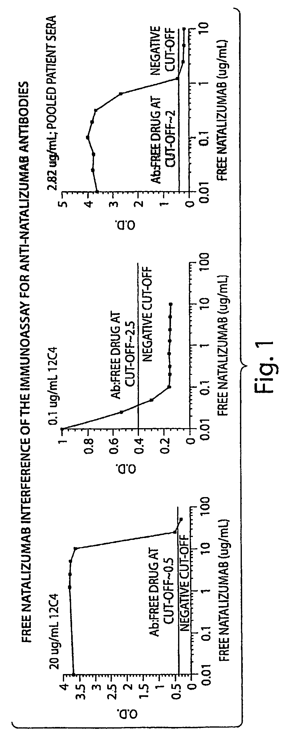
[0119]The tests included the use of a high affinity mAb as a positive control. The high affinity mAb detected binding antibodies (e.g. antibodies that bound natalizumab) that were present in biological samples from subjects. The negative cut-off level was determined to the lowest concentration that returned acceptable accuracy / precision (25% / 25%); 50 ng / mL in 10% serum. The sensitivity of the mAb was 500 ng / mL in neat (undiluted) serum. With respect to sensitivity, the mAb was dete...
example 3
[0122]A screening assay (as described in Examples 1 and 2) was performed on samples from 625 subjects who had been administered natalizumab. The presence of an immune response to a VLA-4 antibody was examined in patients who had undergone natalizumab administration. Incidence of antibodies to natalizumab in the patients examined was: 91% (569 patients) antibody negative and 9% (56 patients) “binding antibody” positive at any time point with 3% (19 patients) “transiently” positive and 6% (37 patients) “persistently” positive. The transient positive patients had detectable antibodies (at a concentration of >0.5 μg / ml) at a single time point, but negative for antibodies at all other time points. The persistent positive patients had detectable antibodies at two or more time points that were at least 42 days apart, or at a single time point with no follow-up samples tested.
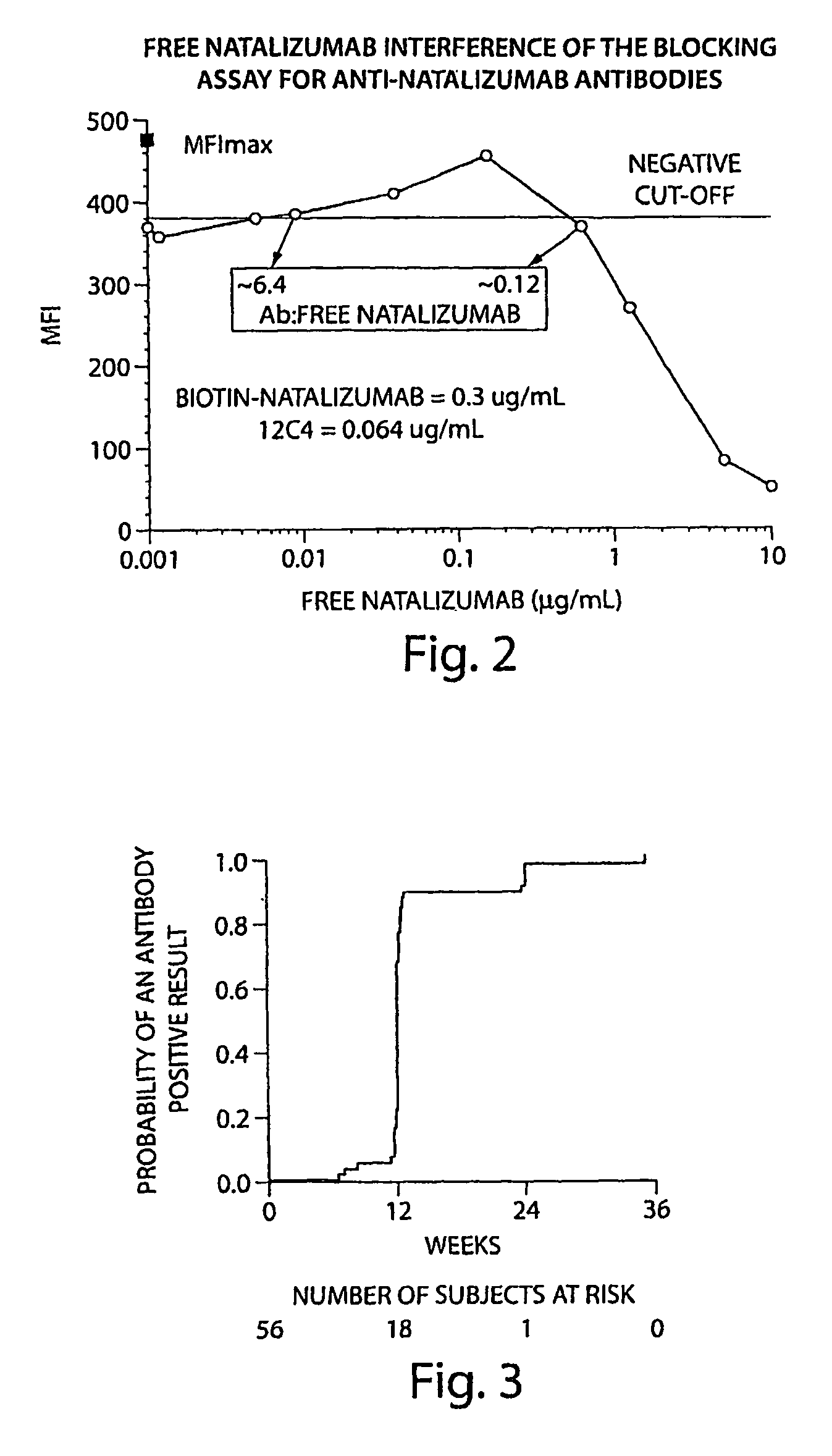
[0123]FIG. 3 shows the time of first positive results in patients who developed any antibodies. Six percent of patie...
PUM
| Property | Measurement | Unit |
|---|---|---|
| time | aaaaa | aaaaa |
| time | aaaaa | aaaaa |
| time | aaaaa | aaaaa |
Abstract
Description
Claims
Application Information
 - R&D Engineer
- R&D Manager
- IP Professional
- Industry Leading Data Capabilities
- Powerful AI technology
- Patent DNA Extraction
Browse by: Latest US Patents, China's latest patents, Technical Efficacy Thesaurus, Application Domain, Technology Topic.
© 2024 PatSnap. All rights reserved.Legal|Privacy policy|Modern Slavery Act Transparency Statement|Sitemap