Ryanodine receptor inhibitors for treatment of T-cell mediated disorders
a technology inhibitors, which is applied in the field of t cell mediated disorders with ryanodine receptor inhibitors, can solve the problems of largely unsatisfactory available therapies for these t cell mediated diseases, and achieve the effect of suppressing its biological activity
- Summary
- Abstract
- Description
- Claims
- Application Information
AI Technical Summary
Problems solved by technology
Method used
Image

Examples
example 1
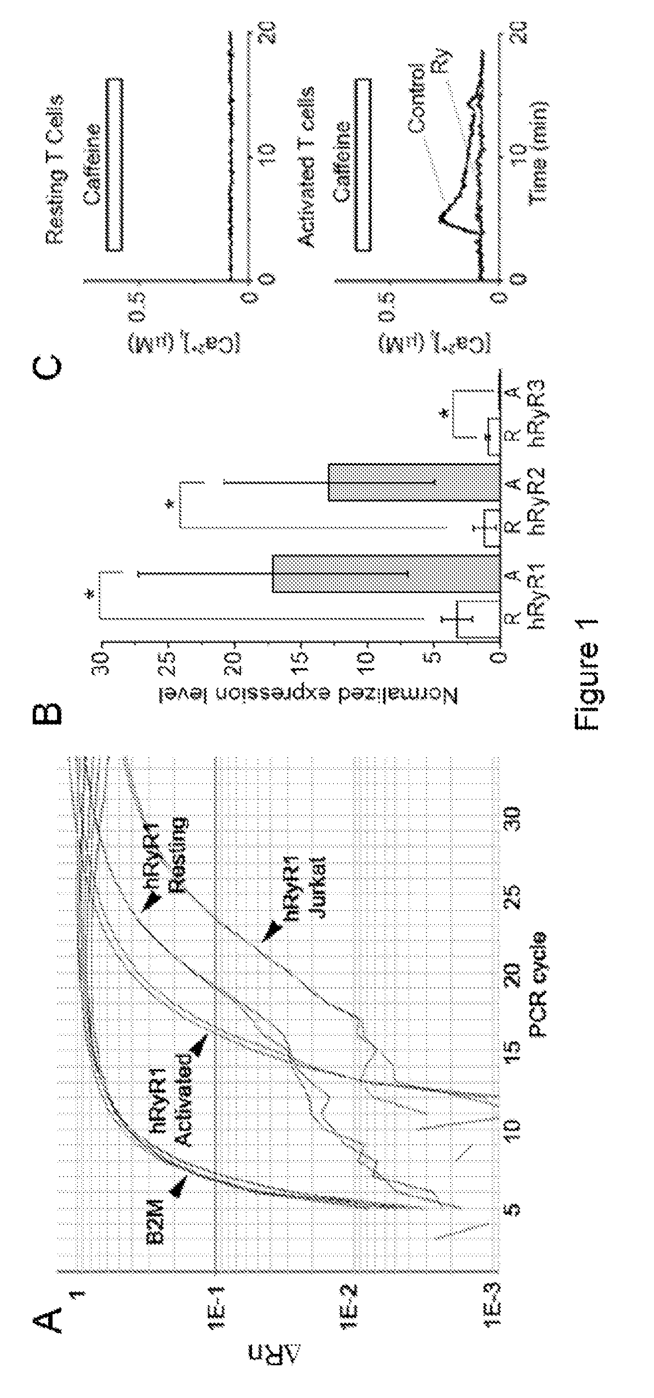
Overexpression of RyR1 Gene Expression in Activated Human T Cells Compared with Resting Human T Cells
Experimental Procedures
[0068]Cell Cultures and Chemicals—
[0069]All studies involving human subjects were performed after approval by UC Davis Institutional Review Board and after all subjects gave written informed consent. Resting CD3+ human lymphocytes were isolated from the peripheral blood of healthy consented adults using RosetteSep™ human lymphocyte enrichment kit (Stem Cell Tech.) following the manufacturer's instructions. Resting T cells were resuspended at 0.5−1×10⁶ cells / ml in RPMI 164 medium supplemented with 10 mM HEPES, 10% fetal bovine serum, 2% L-glutamine, 2% vitamins solution, 1% RPMI amino acids solution, and 0.01% β-mercaptoethanol and maintained in suspension in 5% CO2 at 37° C. Resting T cells were activated on the day of isolation by incubation with either PHA (5-20 μM) or anti-CD3 antibody coated onto the tissue culture plates and soluble anti-CD28 antibody (5 m...
example 2
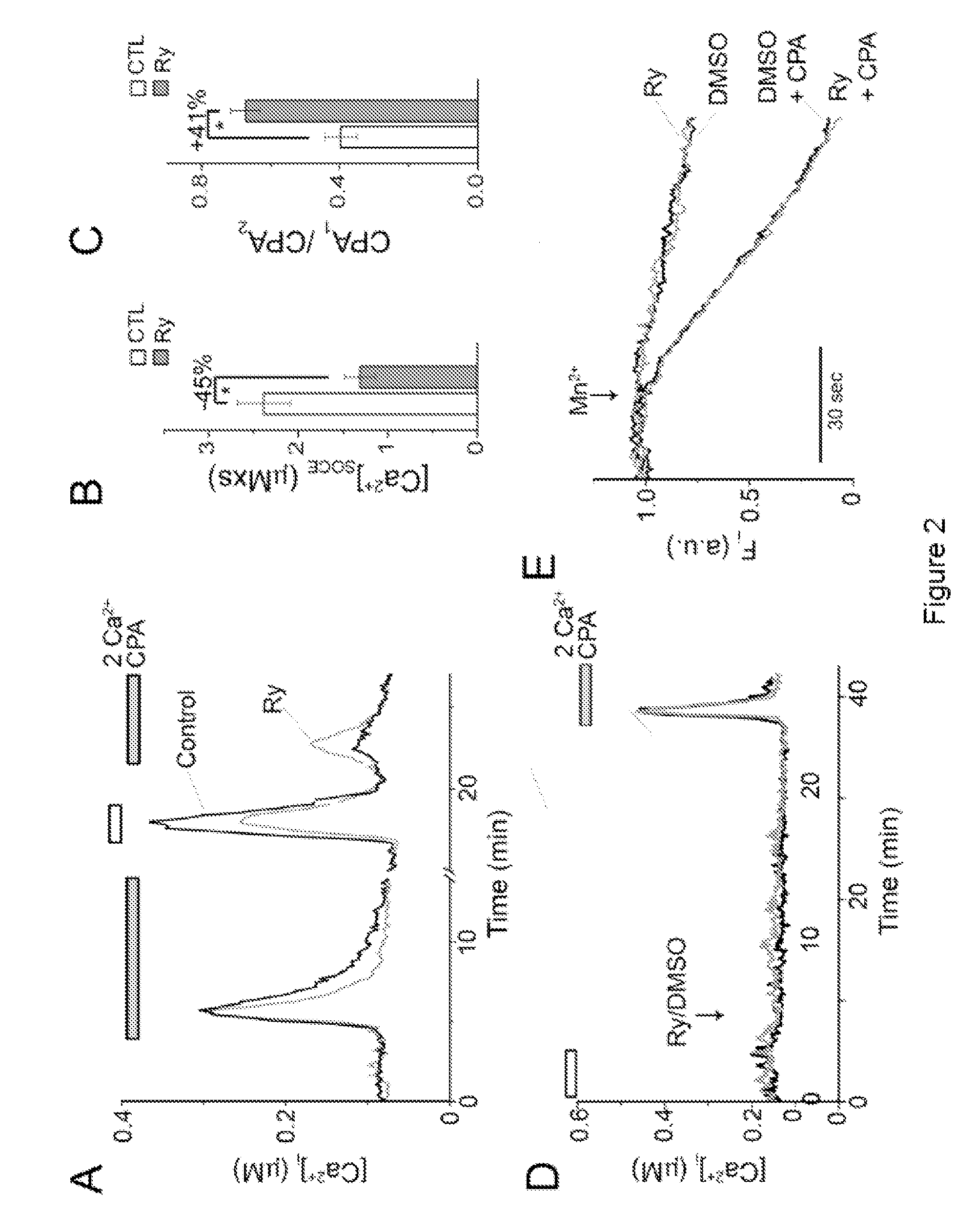
RyR Blockers Ryanodine and Dantrolene Suppress Cytosolic Ca2+ Signaling in T Cells
Experimental Procedures
[0076]Calcium Imaging—
[0077]Cell cultures and chemicals were as described in the Example 1. Before experiment, cells were plated on poly-l-lysine-coated glass-bottom clambers and then loaded with 1 μM fura-2 / AM and pluronic F-127 for 5 min in modified Tyrode solution containing (in mM): 130 NaCl, 5.6 KCl, 1 MgCl2, 2 CaCl2, 10 HEPES, 10 D-glucose; pH 7.3. After washing, cells were incubated for additional 30 min at 37° C. Some cells were incubated in Modified Tyrode solution containing 400 μM Ry, 30 μM dantrolene, or vehicles (methanol or, ethanol, or DMSO) alone for 30 min at 37° C. In nominally Ca2+-free solution, Ca2+ was omitted.
[0078]Fluorescence images were acquired from adherent cells using a SenSys CCD camera (Roper Scientific, Tucson, Ariz.) and a 40× oil immersion Zeiss objective on a Zeiss Axiovert 200 inverted microscope (Thornwood, N.Y.). Lambda DG-4 filter changer (S...
example 3
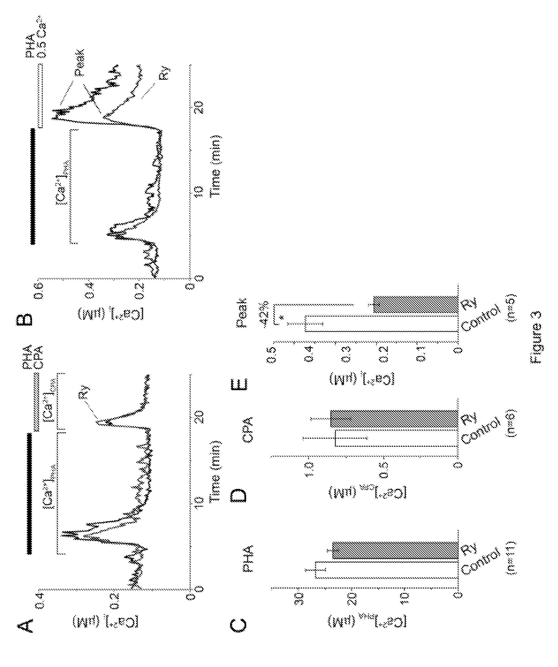
RyR Blockers Ryanodine and Dantrolene Suppress Ca2+-Dependent T Cell Functions
Experimental Procedures
[0095]Cell cultures and chemicals were as described in the Example 1. Proliferation assay—Cell division track assay was conducted using carboxyfluorescein succinimidyl ester (CFSE) proliferation kit (Molecular probes). Stock solution of CFSE (4 mM) was prepared in DMSO. Cells were washed, resuspended in PBS containing 4 μM CFSE at density 1×10⁶ cells / ml, and incubated at 37° C. for 10 min. Labeling was quenched by adding 5× volume of RPMI 1640 culture media containing 10% FBS. After washing 3 times with RPMI 1640+10% FBS, cells were placed into FBS-free cell culture media to abolish cell division and kept overnight in 5% CO2 at 37° C. After overnight incubation, the CFSE-labeled cells were pellet down and fraction of them (˜50000) were fixed with 1% paraformaldehyde in PBS and analyzed by flow cytometry to establish the CFSE fluorescence profile of undivided cells. The remaining CFSE...
PUM
| Property | Measurement | Unit |
|---|---|---|
| excitation wavelengths | aaaaa | aaaaa |
| excitation wavelengths | aaaaa | aaaaa |
| concentration | aaaaa | aaaaa |
Abstract
Description
Claims
Application Information
 - R&D Engineer
- R&D Manager
- IP Professional
- Industry Leading Data Capabilities
- Powerful AI technology
- Patent DNA Extraction
Browse by: Latest US Patents, China's latest patents, Technical Efficacy Thesaurus, Application Domain, Technology Topic.
© 2024 PatSnap. All rights reserved.Legal|Privacy policy|Modern Slavery Act Transparency Statement|Sitemap