Ultrasonic image assistant diagnostic system

A technology of ultrasonic imaging and auxiliary diagnosis, which is applied in the direction of ultrasonic diagnosis, infrasonic diagnosis, ultrasonic/sonic wave/infrasonic diagnosis, etc. It can solve the problems of inability to diagnose diseases and achieve the effect of a good auxiliary diagnostic tool

Inactive Publication Date: 2009-04-01
SHENZHEN LANDWIND IND
View PDF0 Cites 13 Cited by
  • Summary
  • Abstract
  • Description
  • Claims
  • Application Information

AI Technical Summary

Problems solved by technology

[0003] The technical problem to be solved by the present invention is to provide an ultrasonic image-assisted diagnosis system, which overcomes the defect that the existing technology cannot quickly diagnose diseases based on ultrasonic images when encountering difficult or rare cases

Method used


Image

  • Ultrasonic image assistant diagnostic system
  • Ultrasonic image assistant diagnostic system
  • Ultrasonic image assistant diagnostic system

Examples


Embodiment Construction

[0024] Below according to accompanying drawing and embodiment the present invention will be described in further detail:

[0025] 1. Save the real-time image of the ultrasonic diagnostic instrument

[0026] The real-time ultrasonic image in the ultrasonic diagnostic instrument is saved to the disk of the current server through the video acquisition card to become the current static ultrasonic image.

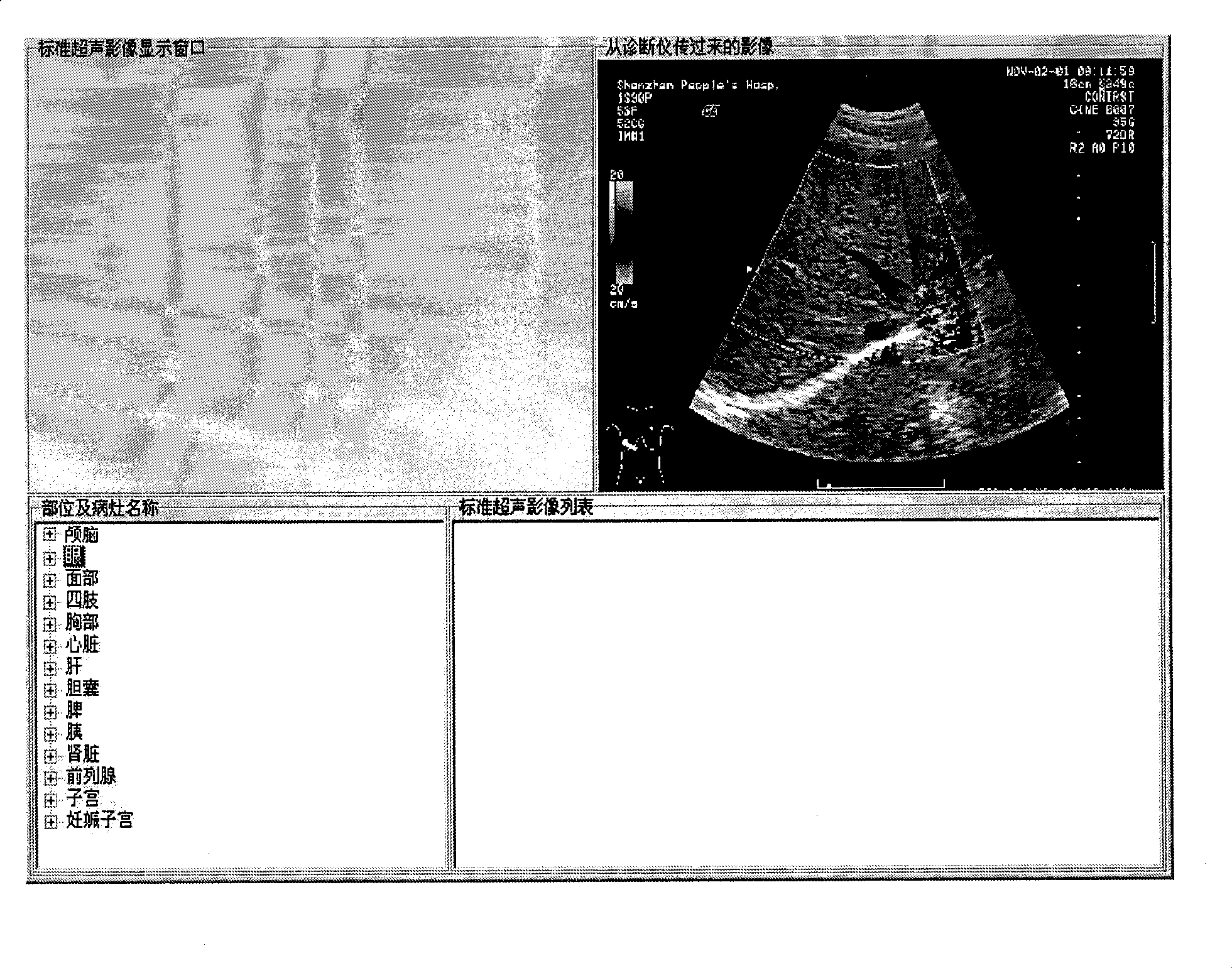
[0027] 2. Generate the main form and load the static ultrasound image

[0028] The steps to generate the main form using the development language Delphi are as follows:

[0029] Create a new project, generate a form, and set the height of the form to 768 and the width to 1024. Call the API function CreateWindow to create a window as the static ultrasound image display window transmitted by the ultrasound diagnostic instrument. The height of the window is 512 pixels and the width is 384 pixels. An image saved on disk is loaded into this Image control.

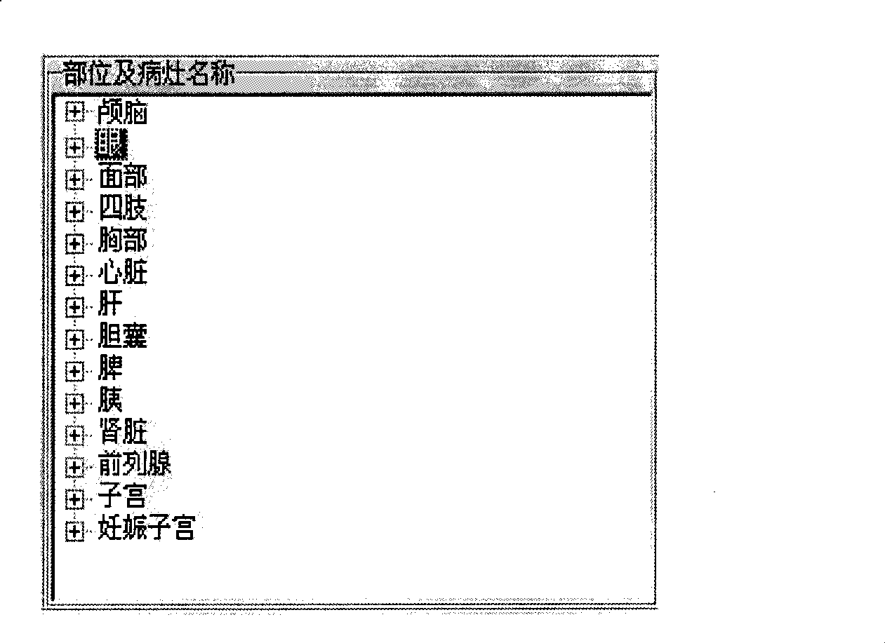
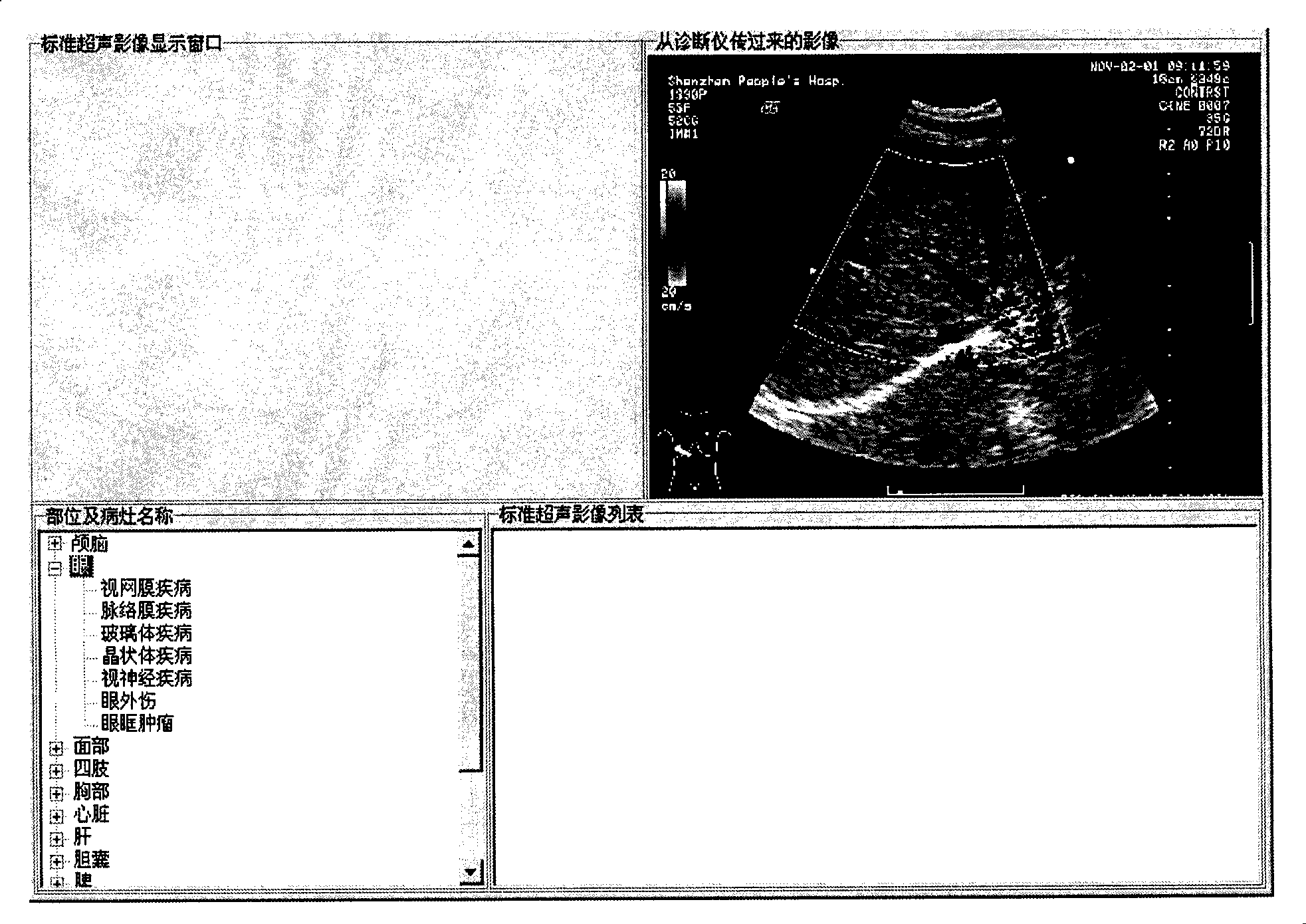
[0030] 3. Create a tree...


PUM


Abstract

The invention discloses an ultrasound image aided diagnosis system which comprises a display interface, a focus name catalog structure which is arranged on the display interface, a standard ultrasound image display area which is arranged on the display interface and is corresponding to a focus name, and a static ultrasound image display area which is arranged on the display area. After the system starts, the static ultrasound image display area displays a static ultrasound image; after the focus name in the focus name catalog structure is clicked, the standard ultrasound image display area displays a standard ultrasound image which is corresponding to the focus name. The ultrasound image aided diagnosis system compares the standard ultrasound image which is corresponding to the focus name and the static ultrasound image which is bypassed by an ultrasound diagnostic unit, and assists an ultrasound doctor to look for characteristic presentation, thus making the right diagnosis on difficult or rare clinical cases and providing good aided diagnosis tool for ultrasound doctors.

Description

technical field [0001] The invention relates to the technical field of ultrasonic imaging, in particular to an ultrasonic image auxiliary diagnosis system. Background technique [0002] With the popularization and application of ultrasound, the majority of primary hospitals in my country basically have an ultrasound department. Its wide range of uses and effectiveness make it an indispensable tool in the field of clinical medicine. However, there are only one or two doctors in the ultrasound department of many primary hospitals. When encountering difficult or rare cases in daily diagnosis, they are often unable to discuss with colleagues and seek advice from experts, so that they cannot draw corresponding conclusions on the examination. Moreover, since ultrasound is a new specialty, the vast majority of existing ultrasound doctors have transferred from clinical practice, and very few of them have graduated from imaging majors. Therefore, they have little understanding of man...

Claims


Application Information

Patent Timeline
Patent Type & Authority Applications(China)
IPC IPC(8): A61B8/00
Inventor 李胜利旷文维
Owner SHENZHEN LANDWIND IND
Why Eureka
Try Eureka