Synchronization for multi-directional ultrasound scanning

A technology of ultrasonic scanning and transducer, applied in the field of ultrasonic scanning, can solve problems such as inability to observe anatomical structures and difficulty in comparing images

Inactive Publication Date: 2011-05-25
SIEMENS MEDICAL SOLUTIONS USA INC
View PDF7 Cites 29 Cited by
  • Summary
  • Abstract
  • Description
  • Claims
  • Application Information

AI Technical Summary

Problems solved by technology

However, the FOV is also limited by the transducer design, so the entire anatomy of interest cannot be observed
Transducers may be positioned at other locations for scanning other areas, but movement of the fetus or within an area may make it difficult to compare images from different scans

Method used


Image

  • Synchronization for multi-directional ultrasound scanning
  • Synchronization for multi-directional ultrasound scanning
  • Synchronization for multi-directional ultrasound scanning

Examples


Embodiment Construction

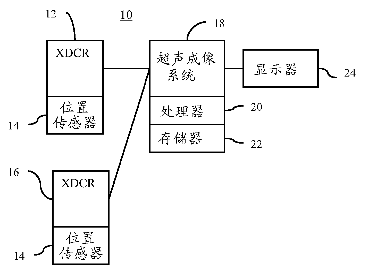
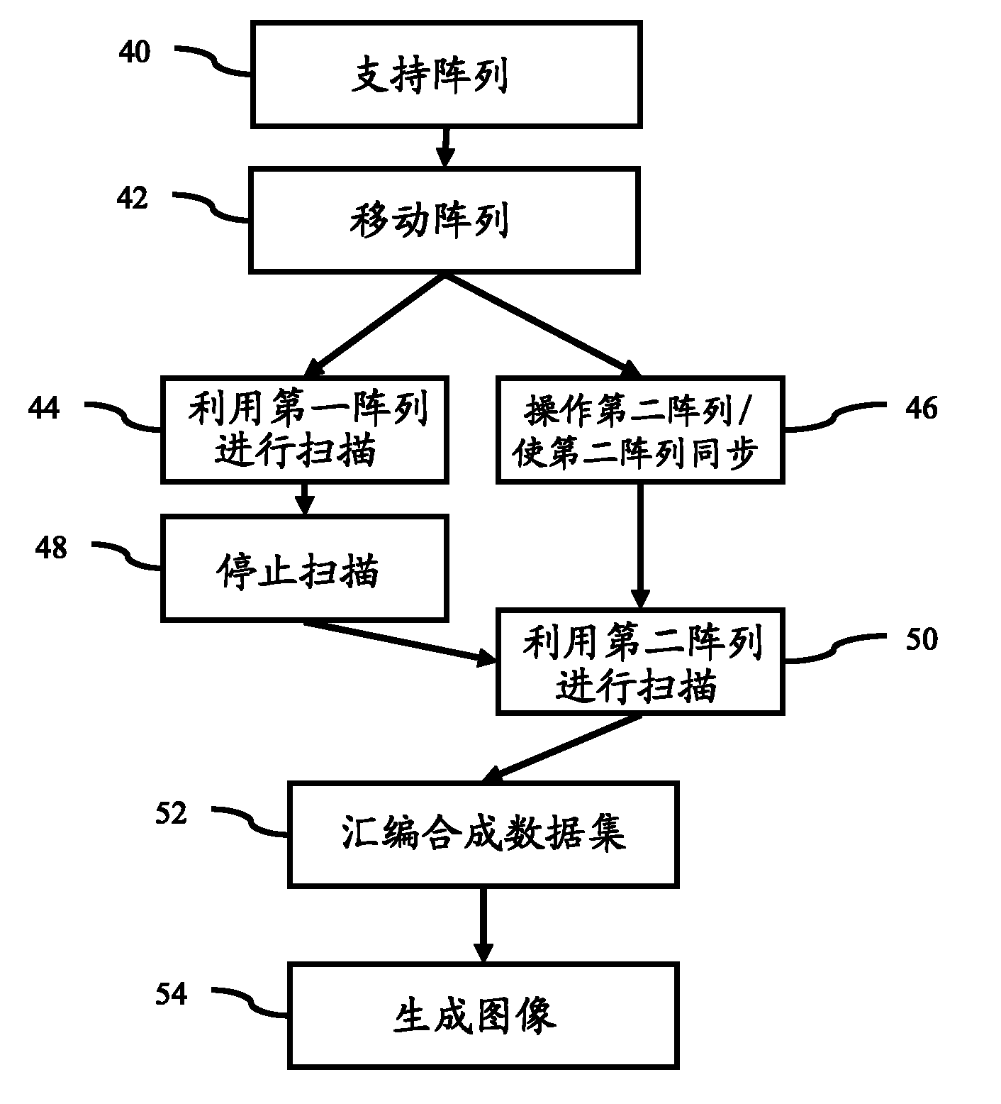
[0013] Synchronization of two or more mechanical oscillating transducers can allow for faster acquisition. Large FOVs can be synthesized using spatially encoded information from each transducer. Multiple transducers with overlapping fields of view are used to compound a volume or plane representing the enlarged field of view.

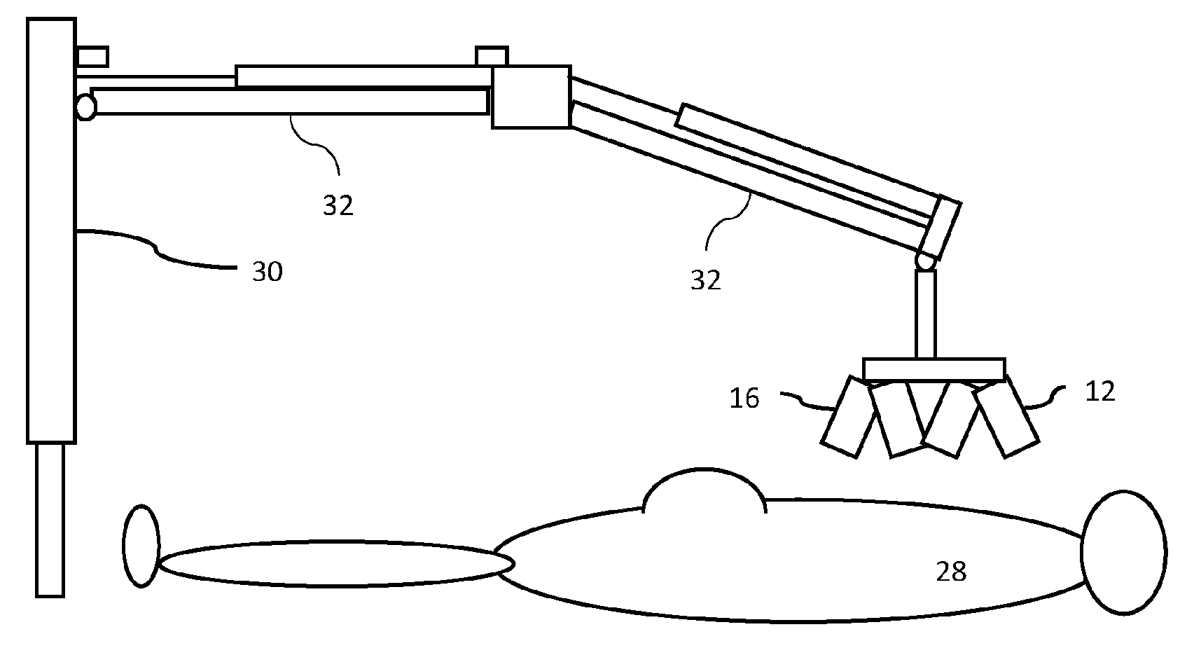
[0014] Composite information can be used for quantification and / or imaging. For example, providing obstetrical imaging. Whole fetal scans can be provided. Ultrasound visualization of other large anatomical structures can be provided using transducer arrays. The transducer array consists of independently arranged transducers with overlapping fields of view (FOV). Each transducer can be addressed sequentially or simultaneously in the transducer array so that a composite large FOV volume can be combined. Synthesis of the resulting volumes is performed using knowledge of the geometry and orientation of the individual transducers and / or using image proc...


PUM


Abstract

Multi-directional ultrasound scanning (44, 50) is synchronized (46). A plurality of wobbler arrays (12, 16) are used sequentially. To limit artifacts caused by motion, the sequential operation is synchronized (46). While a first wobbler array is scanning (44), a second wobbler array is moving or active. Once the first wobbler array completes a scan or portion of the scan, the second wobbler array begins the scan (50) without waiting for initiation of the wobbling. The position of the second array may alternatively or additionally be synchronized with the first array or the end of the scan of the first array. The data from the different scans may represent overlapping volumes, so may be combined (52) to form an extended field of view.

Description

technical field [0001] This embodiment involves ultrasound scanning. In particular, these embodiments relate to scanning in different directions. Background technique [0002] Traditional ultrasonography is performed using a single hand-held transducer. A transducer acquires planar information within a field of view (FOV) limited by the design of the transducer. There are many clinical applications, including fetal imaging, for which this method does not enable visualization of the entire anatomical structure of interest. Instead, multiple independent observations are often required to fully visualize the anatomy of interest. The sonographer moves the handheld transducer to different locations and acquires data independently at each location. Separate images are generated from the data acquired at each location. [0003] Volumetric information can be obtained with the aid of a hand-held transducer. For example, a wobble transducer moves the array mechanically for elect...

Claims


Application Information

Patent Timeline
Patent Type & Authority Applications(China)
IPC IPC(8): A61B8/00
CPCA61B8/54A61B8/5215A61B8/4461A61B8/4477A61B8/461A61B8/4472A61B8/523A61B8/4218A61B8/483A61B8/5253G16H50/20
Inventor R.拉泽布尼克
Owner SIEMENS MEDICAL SOLUTIONS USA INC
Why Eureka
Try Eureka