Cardiac muscle biopsy system with electrophysiology standard measurement and three-dimensional positioning functions

A myocardial biopsy and three-dimensional positioning technology, applied in diagnostic recording/measurement, medical science, sensors, etc., can solve the problems of atrioventricular block, easy damage, and easy wrong clamping of myocardium, so as to reduce the risk of cardiac perforation, The effect of improving the positive detection rate and positive detection rate

Active Publication Date: 2012-02-29
汤日波 +2
View PDF3 Cites 10 Cited by
  • Summary
  • Abstract
  • Description
  • Claims
  • Application Information

AI Technical Summary

Problems solved by technology

However, the myocardial biopsy under the guidance of X-rays has the following problems: (1) It is easy to damage the conduction system such as the His bundle and the right bundle branch of the cardiac septum. Failure, if it causes permanent damage to the His bundle, it will also lead to complete atrioventricular block, which needs to be implanted with a pacemaker; (2) It is easy to damage valve tissues such as chordae, which can lead to valvular regurgitation and long-term valvular regurgitation Increased heart burden leads to heart enlargement, heart failure, and arrhythmia; (3) It is difficult to judge whether the biopsy forceps are closely attached to the myocardium, resulting in heart perforation and heart tamponade, which can lead to death if not treated in time. Kober et al. reported that right ventricular biopsy The incidence rate of perforation is as high as 10%, and half of the patients with cardiac perforation need open chest rescue [Kaltenbach M, Loogen F, Olsen EGJ (translated by Hu Gengming et al). Cardiomyopathy and myocardial biopsy. Jinan: Shandong Science and Technology Press, 1981 :26-32];( ) It is easy to accidentally clamp the normal myocardium instead of the diseased myocardium, resulting in missed detection

Method used


Image

  • Cardiac muscle biopsy system with electrophysiology standard measurement and three-dimensional positioning functions
  • Cardiac muscle biopsy system with electrophysiology standard measurement and three-dimensional positioning functions
  • Cardiac muscle biopsy system with electrophysiology standard measurement and three-dimensional positioning functions

Examples


Embodiment Construction

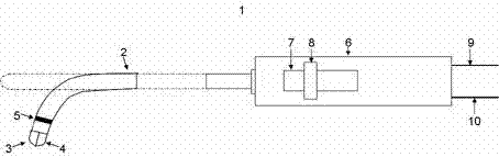
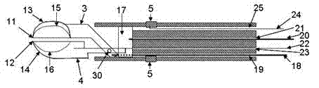
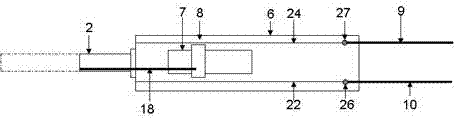
[0026] At least in some embodiments, a myocardial biopsy system with electrophysiological mapping and three-dimensional positioning functions can avoid the shortcomings of low positive detection rate and high complication rate of existing devices. figure 1 A biopsy forceps 1 according to at least some embodiments is shown. The biopsy forceps 1 includes a catheter 2 (diameter 2-5mm). figure 2 The metal clamps 3 and 4 are shown connected to the distal end of the catheter 2. The metal clamps 3 and 4 are connected by a conductive metal shaft 30, the metal clamp 4 is connected with the metal wire 22, and the metal clamps 3 and 4 constitute a remote mapping electrode. The proximal ends of the metal clamps 3 and 4 are ring electrodes 5. The ring electrode 5 is connected to the metal wire 24, and the ring electrode 5 is a proximal mapping electrode. The distal mapping electrode composed of metal clamps 3 and 4 can be used as a monopolar electrode for mapping, or can be used for bipol...


PUM


Abstract

The invention relates to a cardiac muscle biopsy system with electrophysiology standard measurement and three-dimensional positioning functions. The cardiac muscle biopsy system comprises a conduit of which the far end has an adjustable bending degree, a pair of forceps at the far end of the conduit, an operating handle at the near end of the conduit, a tail wire connected with the handle and an electrophysiology standard measurement and three-dimensional positioning device, wherein the pair of forceps at the far end of the conduit are made of metal with high electrical conductivity so as to form a far end standard measurement electrode; the near ends of the metal forceps are provided with a plurality of annular electrodes which are made of the metal with the high electrical conductivity;the metal forceps and the annular electrodes are connected to an electrophysiology standard measurement and three-dimensional positioning system by the tail wire; and the electrophysiology standard measurement and three-dimensional positioning system comprises an electrical signal processing device, a positioning signal processing device, a work station and an output device. By the system, normaland abnormal cardiac muscles are identified through electrophysiology standard measurement, the success rate of biopsy is increased, and complicating diseases are reduced.

Description

Technical field [0001] The invention relates to a myocardial biopsy system, in particular to a myocardial biopsy system with electrophysiological mapping and three-dimensional positioning functions. Background technique [0002] Myocardial tissue biopsy is the gold standard for the diagnosis of cardiomyopathy such as myocarditis, rejection after heart transplantation, and arrhythmogenic right ventricular cardiomyopathy. Myocardial biopsy forceps are instruments used to take tissue samples from the heart forceps of animals or patients. The myocardial biopsy forceps currently used in clinical practice is a flexible catheter that can be manipulated, including a pair of distal end clamps, a proximal control handle, and a catheter connected to the proximal and distal ends. In actual operation, the biopsy forceps are delivered into the heart through the venous blood vessels, and the distal end of the biopsy forceps is placed against the atrium or ventricle under the guidance of X-ray...

Claims


Application Information

Patent Timeline
Patent Type & Authority Applications(China)
IPC IPC(8): A61B5/0488A61B10/02
Inventor 不公告发明人
Owner 汤日波
Why Eureka
Try Eureka