A Segmentation Method of Symmetrical Organs in Medical Images

A medical image and symmetry technology, applied in the field of medical image processing and computer vision, can solve problems such as not considering the symmetry properties of organs

Inactive Publication Date: 2015-12-09
TIANJIN UNIV
View PDF4 Cites 0 Cited by
  • Summary
  • Abstract
  • Description
  • Claims
  • Application Information

AI Technical Summary

Problems solved by technology

This patent has a good performance in terms of segmentation speed and accuracy, but this patent does not consider the symmetrical properties of organs

Method used


Image

  • A Segmentation Method of Symmetrical Organs in Medical Images
  • A Segmentation Method of Symmetrical Organs in Medical Images
  • A Segmentation Method of Symmetrical Organs in Medical Images

Examples


Embodiment Construction

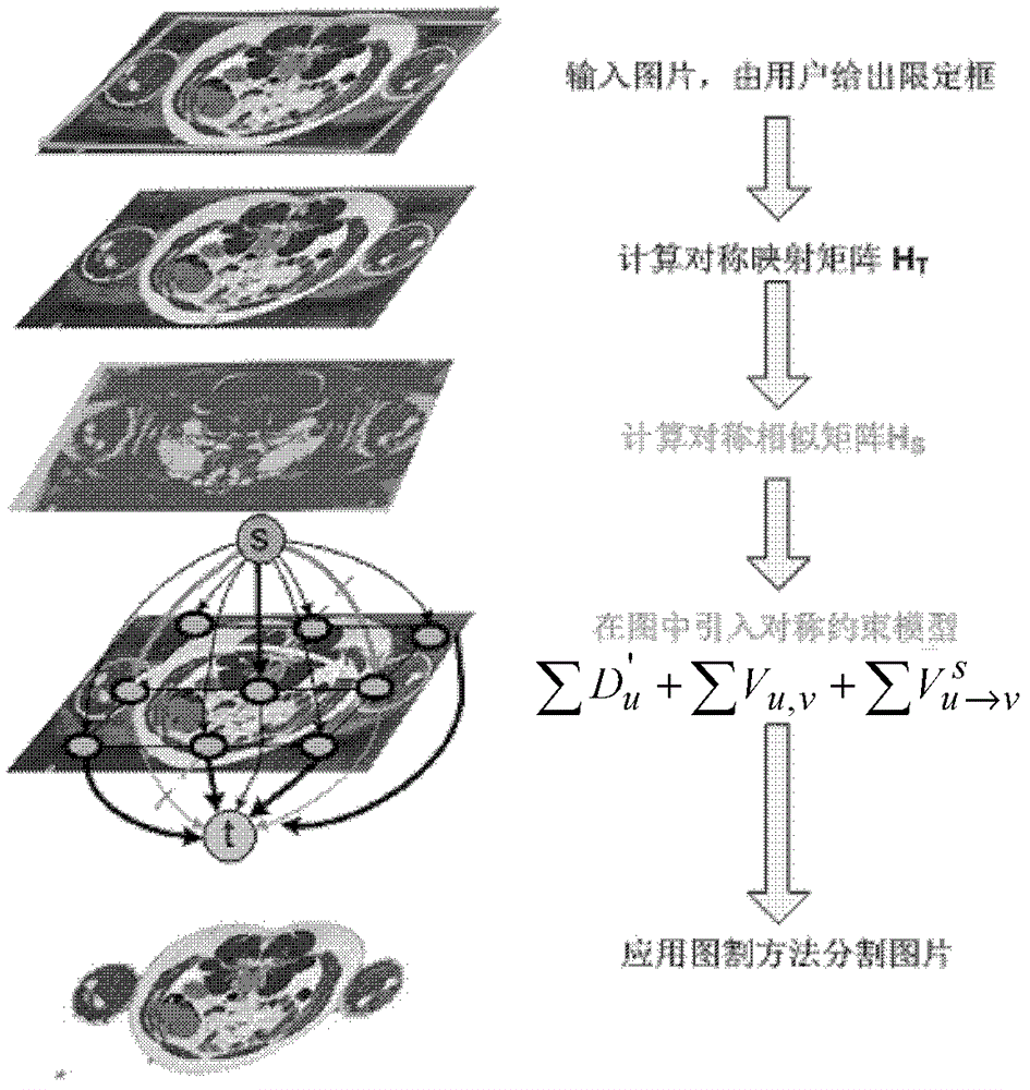
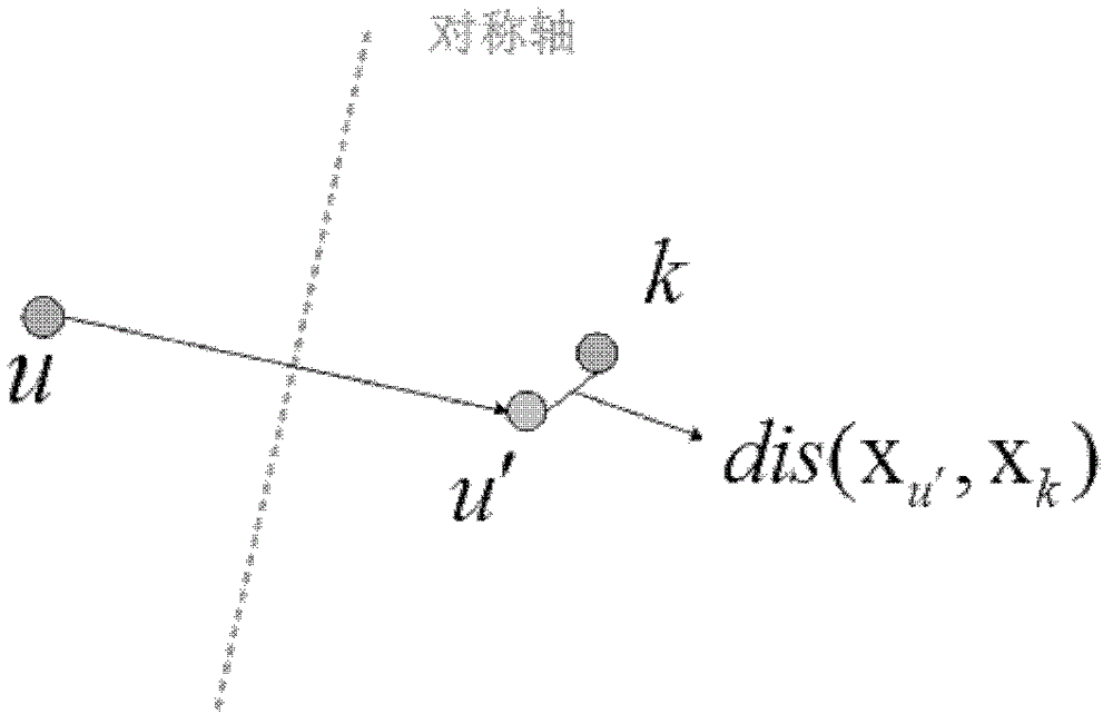
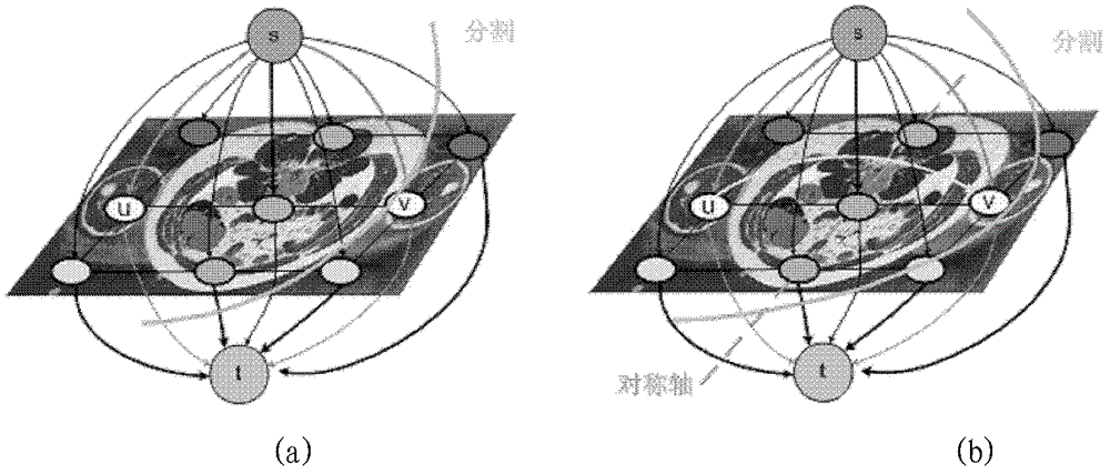
[0051] The invention aims at the problems existing in the traditional medical image segmentation method, and introduces high-level symmetry constraints into the low-level image segmentation method. figure 1 The framework of this method is shown: first, the user gives a bounding box to the input image; then the symmetric mapping matrix is ​​obtained through the symmetry detection algorithm; then the symmetric similarity matrix is ​​introduced to strengthen the symmetric mapping; finally, the symmetric constraint model is introduced in the figure, and through the figure The cut method divides the picture.

[0052] The main contribution of the present invention is to use the symmetric properties of organs to improve the segmentation effect of organs. The symmetric constraint model proposed by the present invention has submodularity [4], and can be solved in polynomial time through the graph cut method.

[0053] There are five steps in the image segmentation method based on the ax...


PUM


Abstract

The invention relates to the technical fields of medical image processing and computer vision and provides a segmentation method of symmetrical organs in a medical image. Symmetrical properties of organs and problems of low speeds and low accuracies are considered in the segmentation method. The technical scheme includes that the segmentation method of the symmetrical organs in the medical image includes the following steps of firstly, confirming a bounding box of the image; secondly, detecting a symmetry axis and calculating symmetrical mapping matrixes; thirdly, obtaining symmetrical similar matrixes; fourthly, introducing symmetrical constraint models to the image; and fifthly, solving an energy equation by means of an image segmentation method to obtain a final segmentation result. The segmentation method of the symmetrical organs in the medical image is mainly applied to medical image processing.

Description

technical field [0001] The invention relates to the technical fields of medical image processing and computer vision, in particular to a method for segmenting symmetrical organs in medical images. Background technique [0002] Digital image processing refers to the process of converting image signals into digital signals and processing them with a computer. With the in-depth development of image processing technology, a great achievement of digital image processing is its application in medical images. In a medical image, people are often only interested in some of the organs, which usually occupy a certain area and are different from adjacent images in some characteristics (such as grayscale, contour, color and texture, etc.) . The differences in these properties can be so pronounced or so subtle that they are not noticeable to the naked eye. Medical image segmentation is one of the prerequisites for its recognition and understanding. The quality of image segmentation di...

Claims


Application Information

Patent Timeline
Patent Type & Authority Patents(China)
IPC IPC(8): G06T7/00
Inventor 操晓春李思远付华柱
Owner TIANJIN UNIV
Why Eureka
Try Eureka