Image optimization method by blending of ultrasound fundamental wave and harmonic wave

An optimization method and fundamental wave technology, applied in ultrasonic/acoustic/infrasonic diagnosis, acoustic diagnosis, infrasonic diagnosis, etc., can solve problems such as low signal-to-noise ratio

Active Publication Date: 2013-07-17
SASET CHENGDU TECH LTD
View PDF6 Cites 13 Cited by
  • Summary
  • Abstract
  • Description
  • Claims
  • Application Information

AI Technical Summary

Problems solved by technology

[0010] The purpose of the present invention is to provide an image optimization method for medical ultrasonic fundamental wave and harmonic fusion, which optimizes the quality of ultrasonic images and overcomes the problems caused by factors such as low detail resolution and contrast resolution of ultrasonic fundamental wave images, artifacts, and gratings. Low image quality and low signal-to-noise ratio caused ...

Method used


Image

  • Image optimization method by blending of ultrasound fundamental wave and harmonic wave
  • Image optimization method by blending of ultrasound fundamental wave and harmonic wave
  • Image optimization method by blending of ultrasound fundamental wave and harmonic wave

Examples


Embodiment Construction

[0051] The technical solutions of the present invention will be further described below in conjunction with the accompanying drawings and specific embodiments.

[0052] An image optimization method for medical ultrasonic fundamental wave and harmonic fusion, comprising the following steps:

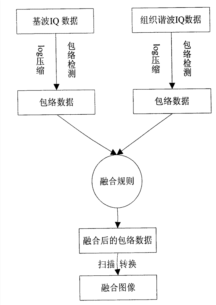
[0053] 1) Obtain fundamental and harmonic envelope data

[0054] The probe alternately transmits at a center frequency of 2f 0 and f 0 signal, for center frequency 2f 0 The signal, directly adopt the center frequency of 2f 0 Fundamental wave signal, through quadrature modulation, low-pass filtering, envelope detection, log compression processing to obtain the envelope signal, the center frequency is f 0 signal, the probe receives a center frequency of 2f 0 The signal of the high-pass filter is used to filter out the center frequency f 0 The fundamental component of , the reserved frequency is 2f 0 The harmonic component of the harmonic component, and then through quadrature modulati...


PUM


Abstract

The invention discloses an image optimization method by blending of ultrasound fundamental wave and harmonic wave. Based on a method of covariance coefficient of texture analysis and Yin Yang normalization, the invention combines the logarithmic compressed ultrasound fundamental wave and the texture harmonic wave, and then a blending image is obtained through scanning and transformation, so that the advantages of both can be utilized and the image quality can be improved.

Description

technical field [0001] The invention belongs to the technical field of medical ultrasonic imaging and display, and relates to an image optimization method for medical ultrasonic fundamental wave and harmonic fusion. Background technique [0002] Medical ultrasound imaging technology is one of the most commonly used clinical medical effects. Because of its safety, real-time, painless, radiation-free, and low price, it is widely used in clinical examination and diagnosis, and is highly favored by medical workers and patients. welcome. B-mode ultrasound fundamental wave imaging FI (fundamental image) is the most basic and most commonly used imaging technology. It can provide the anatomical structure of organs for clinical diagnosis and is widely used in various departments, such as: abdominal organ diagnosis, monitoring fetal status in obstetrics and Diagnosis of heart valves, etc. However, B-mode ultrasound imaging technology also has many limitations. Compared with other eq...

Claims


Application Information

Patent Timeline
IPC IPC(8): A61B8/00
Inventor 李国梁刘西耀刘东权
Owner SASET CHENGDU TECH LTD
Why Eureka
Try Eureka