Imaging method for early breast tumor ultra wide band microwave detection

A microwave detection and imaging method technology, applied in the field of biomedical detection, can solve the problems of human radiation damage, high cost, and low imaging contrast

Inactive Publication Date: 2013-09-18
TIANJIN UNIV
View PDF0 Cites 14 Cited by
  • Summary
  • Abstract
  • Description
  • Claims
  • Application Information

AI Technical Summary

Problems solved by technology

Currently commonly used detection methods for early breast cancer include mammography, ultrasound imaging technology, computed tomography, nuclear magnetic resonance imaging technology, thermal imaging detection, etc., but many methods have certain shortcomings, such as radiation damage to the human body, Low imaging contrast, high cost, etc.

Method used


Image

  • Imaging method for early breast tumor ultra wide band microwave detection
  • Imaging method for early breast tumor ultra wide band microwave detection
  • Imaging method for early breast tumor ultra wide band microwave detection

Examples


Embodiment Construction

[0031] The invention implements the detection of early breast tumors by using a beamforming imaging algorithm on the received signals of the antenna in the ultra-broadband microwave early breast tumor detection system. The present invention will be described below in conjunction with the accompanying drawings and embodiments.

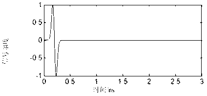
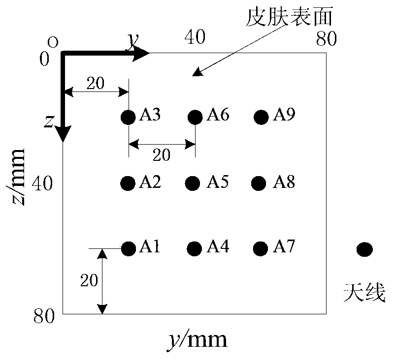
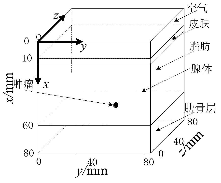
[0032] figure 1 and figure 2 are the antenna array structure and the breast tissue model adopted by the detection system, respectively. In order to meet the requirements of imaging resolution and detection depth, a first-order derivative Gaussian signal with a center frequency of 5GHz and a bandwidth of 10GHz is used. The signal waveform is as follows: image 3 shown. The specific implementation process is as follows:

[0033] 1. Arrange the antenna array directly above the breast model (a phantom is established in this embodiment, which is equivalent to the breast to be tested in the actual microwave test). Select one of the antennas as the trans...


PUM


Abstract

The invention belongs to the technical field of biomedical detection, and relates to an imaging method for early breast tumor ultra wide band microwave detection. The method comprises the steps of performing time delay displacement processing to all detection signals of an antenna array, arranging amplitudes of all groups of signals emitted by an identical transmitting antenna and received by different receiving antennas into a matrix and solving a covariance matrix, performing weighting addition processing to all detection signals of all transmitting antennas, ensuring that weights corresponding to each group of signals are multiplied by the group of detection signals and are added to the group of detection signals, so as to obtain one group of weighting tumor signals, performing windowing processing to the weighting tumor signals, solving squares of all amplitudes in a tumor reflected signal interval and adding the squares, taking a result as a reflection energy value, solving an energy value of a point r, performing smoothing processing to the energy vale of the point r, so as to obtain the weighting energy value of the point r, and performing scanning imaging to a whole breast tissue zone, so as to obtain an energy value distribution diagram inside a breast. The method has good anti-noise capacity.

Description

Technical field [0001] The invention belongs to the technical field of biomedical detection, and relates to a microwave detection and imaging method. Background technique [0002] Breast cancer is the malignant tumor disease with the highest incidence rate in women, and its mortality rate ranks first in the mortality rate of malignant tumors in women. The diagnosis of early breast tumors is undoubtedly of decisive significance for improving the treatment rate of breast disease and the long-term survival rate of patients. Currently commonly used detection methods for early breast cancer include mammography, ultrasound imaging technology, computed tomography, nuclear magnetic resonance imaging technology, thermal imaging detection, etc., but many methods have certain shortcomings, such as radiation damage to the human body, The imaging contrast is low and the cost is high. The principle of ultra-broadband electromagnetic wave detection of breast cancer is that different biol...

Claims


Application Information

Patent Timeline
IPC IPC(8): A61B5/00
Inventor 肖夏徐立王梁
Owner TIANJIN UNIV
Why Eureka
Try Eureka