Polymer magnetic particle composite ultrasonic imaging microsphere

A technology of polymer magnetism and composite ultrasound, which is applied in the medical field, can solve the problems of good biocompatibility, mechanical stability, targeting performance, nanoscale structure, and large size of the ball, etc., to achieve good biological Compatibility, material stability, high mechanical stability effect

Inactive Publication Date: 2015-03-18
CHENGDU LVKE HUATONG TECH
View PDF1 Cites 2 Cited by
  • Summary
  • Abstract
  • Description
  • Claims
  • Application Information

AI Technical Summary

Problems solved by technology

However, this method still has certain limitations, such as hollow SiO 2 The size of the spheres is too large and does not reach the nanoscale structure, which makes it difficult to be used for in vivo imaging, and it is difficult to carry out further connection without functional group modification
Therefore, the contrast agents currently used in clinical practice cannot simultaneously meet the requirements of good biocompatibility, mechanical stability, targeting performance, nanostructure, and functional group modification.

Method used


Image

  • Polymer magnetic particle composite ultrasonic imaging microsphere
  • Polymer magnetic particle composite ultrasonic imaging microsphere
  • Polymer magnetic particle composite ultrasonic imaging microsphere

Examples


Embodiment approach

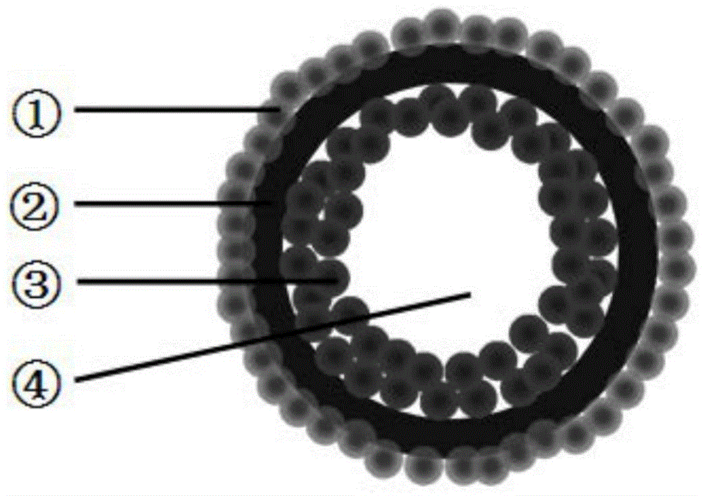
[0038] Polymethacrylic acid-divinylbenzene copolymerized cross-linked polymer is used as the polymer layer ②, and at the same time, the outer layer is adsorbed with silica particles ①, and the inner layer is combined with three-body magnetic nanoparticles ③. The inner layer microspheres are obtained by copolymerizing isopropylacrylamide and polymethacrylic acid, and then the inner layer is copolymerized by polymethacrylic acid and divinylbenzene to obtain double-layer solid nano-polymer microspheres. The carboxyl groups on the surface of the double-layer nano-microspheres can generate hydrogen bonds with the silicon hydroxyl groups on the surface of the silica, so that the silica is hydrolyzed and condensed on the surface of the aggregate to form a shell structure①. At the same time, using the mesoporous structure of silica nanoparticles, the inner core is extracted by the organic solvent method (acetone) to form a hollow polymer-silica double-layer nano-microsphere structure. ...

Embodiment 1

[0043] ① Preparation of core / shell polymer nanospheres

[0044]Step 1: First, monodisperse (PNIPAM-PMAA) copolymer microspheres were prepared by the method of "self-dispersion" polymerization. In a 500mL three-necked round-bottomed reaction flask, 0.2mol of isopropylacrylamide and methacrylic acid, 0.98g of AIBN (azobisisobutylcyanide) and 280mL of butyl acetate were added, and the reaction was carried out at a constant temperature of 70°C for 12 minutes under nitrogen protection. hours, the product was dried in an oven at 55 °C to obtain monodisperse (PNIPAM-PMAA) copolymer microspheres.

[0045] Step 2: Preparation of core / shell polymer microspheres by seeded "self-dispersing" polymerization. 3.65 g of (PNIPAM-PMAA) copolymer microspheres, 0.038 mol of methacrylic acid and divinylbenzene, 0.09 g of AIBN, 100 mL of butyl acetate, and 35 mL of n-hexane were added to a reaction flask, and under nitrogen protection, The reaction was carried out at a constant temperature of 75°...


PUM

PropertyMeasurementUnit
Particle sizeaaaaaaaaaa

Abstract

The invention discloses a polymer magnetic particle composite ultrasonic imaging microsphere. The microsphere has a hollow inorganic / organic / inorganic three-layer structure, wherein a cavity is formed in the center of the microsphere and is filled with air inside; Fe3O4 magnetic particles are formed on the outer layer of the cavity; an intermediate layer is formed by a core / shell polymer nano microsphere obtained through free radical copolymerization of a poly(isopropyl acrylamide)-polymethylacrylic acid random copolymer, polymethylacrylic acid and divinyl benzene; coordinate bonds or ionic bonds can be formed between the copolymer and the Fe3O4 magnetic particles on the inner layer of the copolymer via carboxyl on the inner surface of the microsphere and iron salt, so that the microsphere is stable; an outer layer is formed by silicon dioxide and is connected with the intermediate layer through hydrogen-bond intersection between carboxyl on the surface of the microsphere and silicon hydroxyl on the surface of silicon dioxide. The material has the advantages that the polymer microsphere belongs to nanoparticles; a contrast agent can achieve targeted localization via the Fe3O4 magnetic particles on the inner layer; the silicon dioxide hollow microsphere has good biocompatibility and mechanical stability, and the inside cavity can generate strong echo signals in a contrast mode.

Description

technical field [0001] The invention relates to a contrast agent for medical detection, in particular to a multilayer organic / inorganic composite structure nano microsphere with magnetic function, which belongs to the field of medical technology. Background technique [0002] In recent years, molecular imaging technology has attracted extensive attention because it can provide important information for the diagnosis and monitoring of diseases in living tissues (Campbell R. E., Chang C. J. Curr Opin Chem Biol , 2010, 14(1):1-2.). Among many imaging modes, ultrasound imaging, as a non-invasive biomedical imaging mode, has important research value due to its advantages of safe and timely imaging and diagnosis, low price, lightness and quickness, and wide application. However, the difference in echo signals between different biological tissues often hinders the accuracy of diagnosis. In order to better obtain accurate information of specific tissues, people usually use the meth...

Claims


Application Information

Patent Timeline
IPC IPC(8): A61K49/22C08F220/54C08F220/06C08F212/36C08J9/26B01J13/14
Inventor 不公告发明人
Owner CHENGDU LVKE HUATONG TECH
Why Eureka
Try Eureka