Suspension cell sphere immunofluorescent staining method and staining device

A technique for immunofluorescence staining and suspension cells, which is applied in the field of suspension cell sphere immunofluorescence staining methods and staining devices, can solve the problems of time-consuming and labor-consuming, prolonged antibody incubation time, and long-consuming time, and achieves saving labor and time, saving Experiment cost and the effect of improving the success rate

Active Publication Date: 2018-12-18
GENERAL HOSPITAL OF TIANJIN MEDICAL UNIV
View PDF6 Cites 0 Cited by
  • Summary
  • Abstract
  • Description
  • Claims
  • Application Information

AI Technical Summary

Problems solved by technology

[0005] Chinese patent document CN102288471B discloses a method for immunofluorescent staining of suspension cells. This method is suitable for suspension cells. At least 10 times of cell centrifugation are required to complete the experiment. This method not only consumes time and manpower, but also applies this method to suspension cells. During cell spheroids, due to the different phenotypes of the inner layer cells and the outer layer cells of the cell spheroids, multiple centrifugations can detach the outer layer cells, damage the structure and cell composition of the cell spheroids, and cause the composition of the final test cell spheroids to be different from that before the experimental operation , affecting the accuracy of the experimental results
[0007] Geraldo, S. et al found that immobilizing cell spheres in collagen gel can indeed ensure that the cell spheres will not be lost [8] , but this method still has defects: first, the collagen fibers in the gel will act as a shield, affecting the excitation light intensity and penetration distance of the confocal microscope; second, in order to achieve the same staining effect, it is necessary to The concentration of antibody working solution is doubled or even tripled, and the incubation time of antibody is prolonged, which increases the time consumed and experimental funds [9]
[0008] Guerrero-Cazares, H. et al. studied the use of OCT embedding agent to embed cell spheres to make frozen tissue sections and perform immunofluorescence staining [10] , the disadvantage of this method is that a cryostat is required, and the slices of frozen tissues need to be prepared by professional technical personnel, and it is difficult to find the layer with cell spheres in the process of slicing, and the experimental steps are cumbersome and time-consuming.

Method used


Image

  • Suspension cell sphere immunofluorescent staining method and staining device

Examples


Embodiment 1

[0052] An immunofluorescence staining method for suspension cell spheres, the staining method includes the following steps:

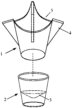
[0053] (I) Combine cell chamber 1 and staining kit 2, take the cell ball suspension and add it to cell chamber 1, let it stand, unplug staining kit 2, and discard the medium in staining kit 2;

[0054] (Ii) Reconstitute cell chamber 1 and staining kit 2. Add 200ul PBS to cell chamber 1 to wash the cell pellets, let stand for 5 minutes, remove staining kit 2, discard PBS in staining kit 2, and repeat the operation three times;

[0055] (Iii) Reconstitute cell chamber 1 and staining kit 2, add 100ul of paraformaldehyde with a mass concentration of 4% to cell chamber 1 and fix for 10 minutes, remove staining kit 2, and discard the liquid in staining kit 2;

[0056] (Iv) Reconstitute cell chamber 1 and staining kit 2. Add 200ul PBS to cell chamber 1 to wash the cell pellets, let stand for 5 minutes, remove staining kit 2, discard PBS in staining kit 2, and repeat th...


PUM


Abstract

The invention discloses a floating sphere immunofluorescent staining method and a staining device, belonging to methods for coloring a sample for test. The staining method comprises the following steps: separation of spheres from a culture medium, fixing by stationary liquid, adding of Triton X-100 transparent cells, closing by confining liquid, adding of an interest protein-resisting primary antibody working solution, adding of a primary antibody-resisting fluorescent secondary antibody working solution, and cell nucleus staining by DAPI dye liquor. The staining device comprises a cell chamber and a staining sleeve part arranged at the outer part of the lower end of the cell chamber in a sleeving way. The floating sphere immunofluorescent staining method has the advantages of high efficiency, simpleness, convenience and the like, an antibody usage amount is reduced, experiment cost is reduced, manpower and time are saved, an accurate and reliable experiment result is obtained on the basis that a fundamental principle that immunofluorescent staining is not changed and specially training experimenters is not needed, sphere loss in an experiment operation process is avoided, an experiment success rate is increased, and the efficiency is improved.

Description

Technical field [0001] The invention belongs to a method for coloring test samples, in particular to a method and a staining device for immunofluorescence staining of suspended cell spheres. Background technique [0002] Immunofluorescence staining technology is a commonly used experimental method of cell and molecular biology. It uses the principle of antigen-antibody reaction to label antibodies with fluorescent substances for antigen localization. It is used to observe the expression of the target protein and whether there is a co-localization or co-expression relationship between the proteins. . [0003] For the more common immunofluorescence staining of adherent cells, it is commonly used and commonly used to "climb" the cells on the surface of the polylysine-coated carrier, and then perform the classic staining operation. However, for the suspension cell spheres commonly used in stem cell research, because it does not adhere to the wall, it is easy to cause the loss of the c...

Claims


Application Information

Patent Timeline
Patent Type & Authority Patents(China)
IPC IPC(8): G01N1/31
CPCG01N1/31
Inventor 杨学军张辰李涛于圣平刘波周星辰
Owner GENERAL HOSPITAL OF TIANJIN MEDICAL UNIV
Why Eureka
Try Eureka