Multi-mode microwave imaging method and system

A microwave imaging and multi-modal technology, applied in medical science, sensors, diagnostic recording/measurement, etc., can solve the problem that the two images cannot be unified, and achieve the effect of complementing the advantages and disadvantages of imaging

Inactive Publication Date: 2016-06-15
YITI INTELLIGENT TECH LTD SHENZHEN CITY
View PDF4 Cites 2 Cited by
  • Summary
  • Abstract
  • Description
  • Claims
  • Application Information

AI Technical Summary

Problems solved by technology

[0004] The technical problem solved by the present invention is to construct a multi-modal microwave imaging method and system to overcome the technical problem that a single detection device in the prior art cannot unify the two images

Method used


Image

  • Multi-mode microwave imaging method and system
  • Multi-mode microwave imaging method and system
  • Multi-mode microwave imaging method and system

Examples


Embodiment Construction

[0022] The technical solutions of the present invention will be further described below in conjunction with specific embodiments.

[0023] Such as figure 1 As shown, the specific embodiment of the present invention is: provide a kind of multimodal microwave imaging method, comprise the steps:

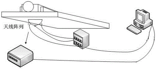
[0024] Microwave scanning: generate microwave broadband pulse signals and microwave single-frequency coherent signals in the area to be measured; receive microwave broadband pulse echo signals and microwave single-frequency echo signals;

[0025] The specific implementation process is as follows: the microwave generating unit generates a microwave broadband pulse signal through the microwave antenna unit to be measured, the microwave broadband pulse signal irradiates the lesion area in the imaging area, produces scattering on the surface of the lesion area, and scans and receives the signal from the lesion with several microwave antenna units. Surface-scattered microwave broadband puls...


PUM


Abstract

The present invention relates to a multi-modal microwave imaging method and system. By mapping the pixel points of one image in the radar detection image and the tomographic image to the other image, the relevant pixel points of the two images are The fusion of the two images is completed when the spatial position is consistent. In the process of diagnosing early cancer patients, different imaging modalities can be used to complement each other. In the present invention, these two technologies are successfully integrated to form a multi-mode microwave breast imaging system to achieve the purpose of complementary imaging.

Description

technical field [0001] The invention relates to an image fusion method and system, in particular to a radar detection image and tomography image fusion method and system. Background technique [0002] In the medical testing process, it usually includes X-ray film, color Doppler ultrasound, magnetic resonance and histological testing. X-ray film has high resolution, but it is accompanied by ionizing radiation and compression of the breast, so it is not suitable for general examination of patients. There is no radioactive damage in color Doppler ultrasound, but it cannot show calcification and burr structures in some cancer patients, and it is greatly affected by the experience of the examiner. The cost of MRI is high, the imaging time is long, and histological examination also requires precise positioning, such as molybdenum target-guided needle insertion, color Doppler ultrasound-guided needle insertion, etc. Therefore, these current detection methods are not suitable for ...

Claims


Application Information

Patent Timeline
Patent Type & Authority Applications(China)
IPC IPC(8): A61B5/05
CPCA61B5/0507
Inventor 张晓峰
Owner YITI INTELLIGENT TECH LTD SHENZHEN CITY
Why Eureka
Try Eureka