
Simulated positioning device and simulated positioning method for intra-operative image guided radiation therapy

A simulated positioning and image-guided technology, applied in radiation therapy, X-ray/γ-ray/particle irradiation therapy, treatment, etc., can solve problems such as lack of dose distribution information, insufficient dose in the target area, and affecting the effect of intraoperative radiotherapy

Active Publication Date: 2016-08-24
CANCER INST & HOSPITAL CHINESE ACADEMY OF MEDICAL SCI
View PDF3 Cites 15 Cited by
  • Summary
  • Abstract
  • Description
  • Claims
  • Application Information

AI Technical Summary

Problems solved by technology

[0003] The current treatment plan for IORT is decided by radiologists based on experience within a short period of time after being on the operating table. Due to the lack of intraoperative three-dimensional (3D) images and dose distribution information, it is impossible to evaluate the three-dimensional dose of the irradiated tissue. The correct choice of parameters (e.g., size, angle, energy, etc.) of the treatment light tube largely depends on the doctor's experience level
This may lead to insufficient dose in the target area or excess dose in important organs, which will affect the effect of intraoperative radiotherapy

Method used


Image

  • Simulated positioning device and simulated positioning method for intra-operative image guided radiation therapy
  • Simulated positioning device and simulated positioning method for intra-operative image guided radiation therapy
  • Simulated positioning device and simulated positioning method for intra-operative image guided radiation therapy

Examples


Embodiment Construction

[0038] Various embodiments according to the present application will be described in detail with reference to the accompanying drawings. Here, it is to be noted that, in the drawings, the same reference numerals are assigned to components having substantially the same or similar structures and functions, and repeated descriptions about them will be omitted.

[0039] Since the treatment light tube and its accessories of intraoperative radiotherapy equipment currently used clinically are made of metal materials, it is impossible to use common imaging equipment in the operating room (for example, C-arm system) to image the patient's intended radiotherapy site. for imaging.

[0040] In view of the above technical problems, in the embodiment of the present application, a simulation positioning device and method for intraoperative image-guided radiotherapy is proposed, which can cooperate with imaging equipment to image the patient's intended radiotherapy site, without unnecessary ...


PUM


Abstract

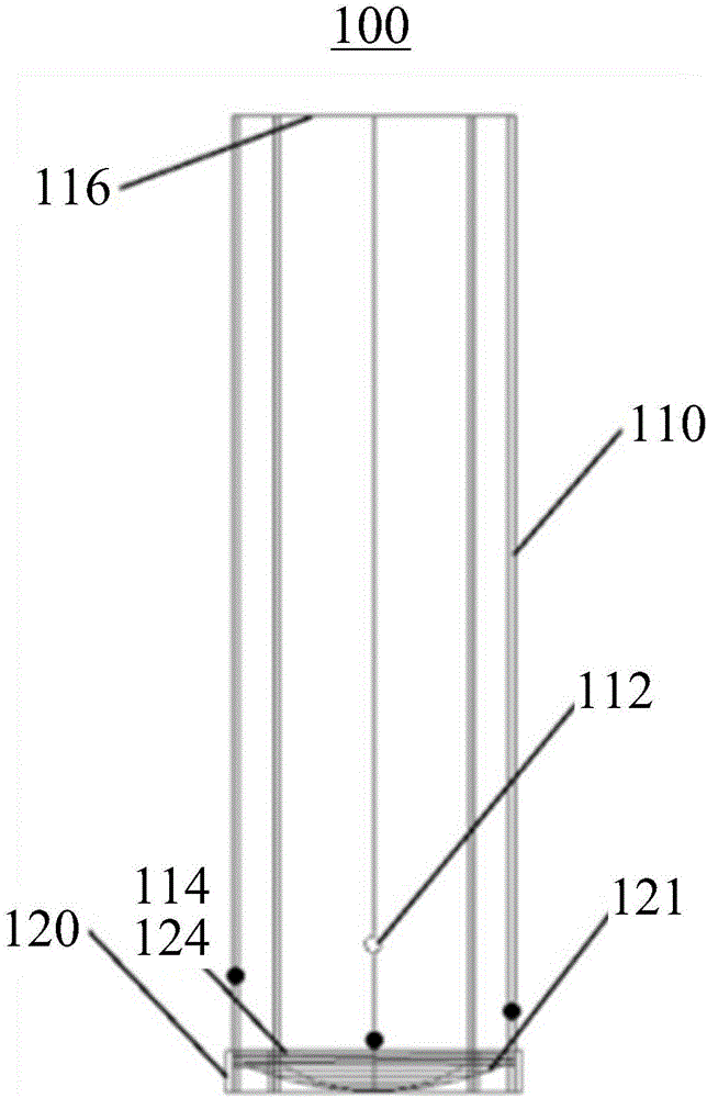
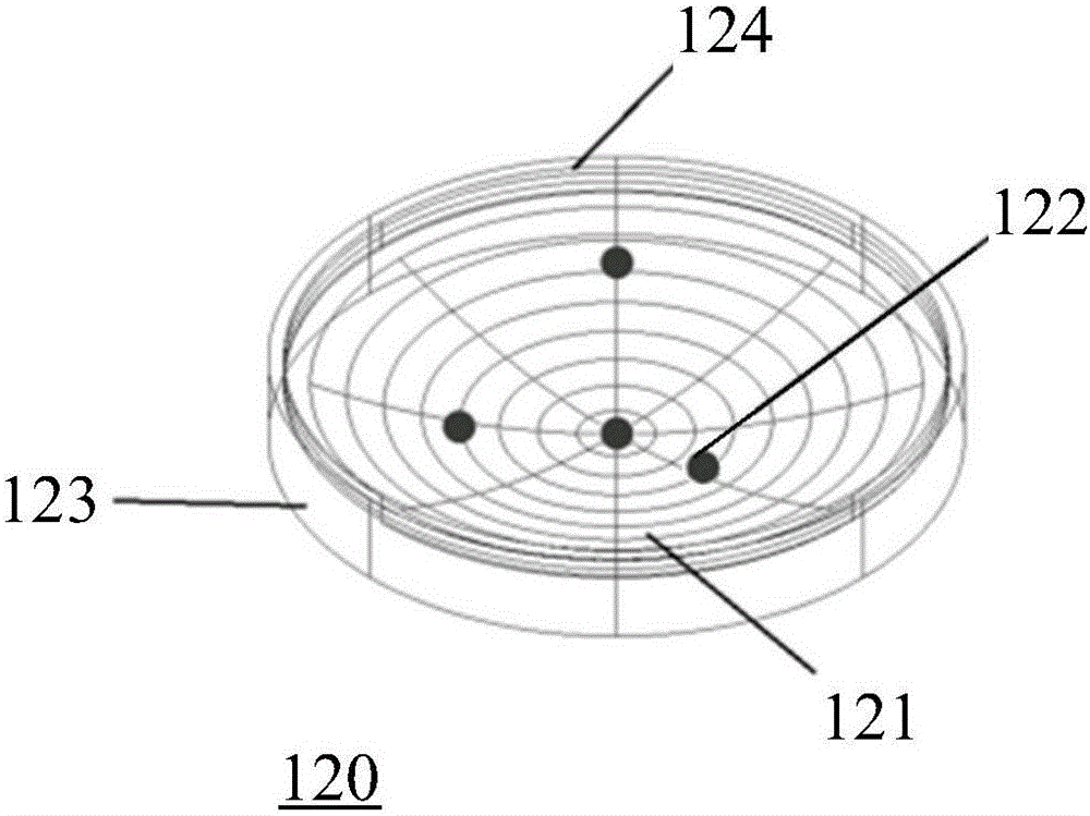
The invention discloses a simulated positioning device and a simulated positioning method for intra-operative image guided radiation therapy. The simulated positioning device comprises a multifunctional light limiting cylinder, and the multifunctional light limiting cylinder is composed of a material which imaging equipment can penetrate to image a body part of a patient about to receive radiation treatment. The multifunctional light limiting cylinder is provided with a positioning mark which is composed of a material different from that of the multifunctional light limiting cylinder so the positioning mark can be distinguished from other areas in display in an image formed by the imaging equipment, and the distinct display of the positioning mark is for positioning the body part about to receive radiation treatment. Therefore, the simulated positioning device disclosed by an embodiment of the invention can cooperate with the imaging equipment to image a body part of a patient about to receive radiation treatment, no unnecessary barrier is produced, and the need of intra-operative image guided radiation therapy is well satisfied.

Description

technical field [0001] The present application generally relates to the field of intraoperative radiotherapy, and more specifically, relates to a simulation positioning device and method for intraoperative image-guided radiotherapy. Background technique [0002] Intra-Operative Radiation Therapy (IORT) is a high-dose radiotherapy under direct vision for visible tumors, tumor bed areas or sites prone to recurrence and metastasis during surgery. The biggest advantage of this technique is that it can push some important organs close to the target area of ​​the patient (ie, the site to be irradiated) out of the radiation field under surgical conditions, thereby reducing their exposure dose. [0003] The current treatment plan for IORT is decided by the radiologist based on experience within a short period of time after being on the operating table. Due to the lack of intraoperative three-dimensional (3D) images and dose distribution information, it is impossible to evaluate the ...

Claims


Application Information

Patent Timeline
Patent Type & Authority Applications(China)
IPC IPC(8): A61N5/10A61N5/00
CPCA61N5/00A61N5/1049
Inventor 戴建荣马攀李明辉陈辛元
Owner CANCER INST & HOSPITAL CHINESE ACADEMY OF MEDICAL SCI
Learn More