Tumor identification method

An identification method and tumor technology, applied in the field of tumor identification, can solve the problems of staging identification, inability to provide a more objective organ tumor identification method, and inability to identify benign/malignant tumors, and achieve the objective effect of the identification method

Active Publication Date: 2017-10-24
SUZHOU INST OF BIOMEDICAL ENG & TECH CHINESE ACADEMY OF SCI
View PDF9 Cites 15 Cited by
  • Summary
  • Abstract
  • Description
  • Claims
  • Application Information

AI Technical Summary

Problems solved by technology

[0005] Although the above two segmentation methods can segment tumors in human organs, they cannot perform benign/malignant identification and malign

Method used


Image

  • Tumor identification method
  • Tumor identification method
  • Tumor identification method

Examples


Embodiment Construction

[0048] The present invention will be further described in detail below in conjunction with the accompanying drawings, so that those skilled in the art can implement it with reference to the description.

[0049] It should be understood that terms such as "having", "comprising" and "including" as used herein do not entail the presence or addition of one or more other elements or combinations thereof.

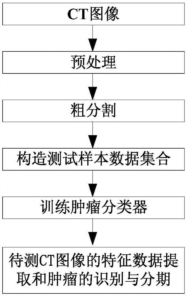
[0050] Such as figure 1 As shown, the present invention provides a method for tumor identification, which comprises the following steps:

[0051] S10, rough segmentation: sequentially perform organ segmentation and blood vessel segmentation on the CT image, and obtain a CT image of the organ without blood vessels.

[0052] S20, constructing a test sample data set: based on the organ CT image, sequentially constructing arterial phase, venous phase, and delay phase tumor region sub-atlases to form a test positive sample data set, and organ normal region sub-atlases to form a test ...


PUM


Abstract

The invention discloses a tumor identification method, which comprises the following steps: rough segmentation: sequentially performing organ segmentation and blood vessel segmentation on a CT image to obtain a CT image of an organ with blood vessel being removed; constructing a test sample data set: based on an the CT image of the organ, sequentially constructing the tumor area sub-graph sets for an arterial phase, a venous phase and a delayed phase to form a test positive sample data set and the organ normal area subgraph set constitutes the test negative sample data set. The training tumor classifier: extracts the characteristic data of the test sample data set, Good/bad identification and staging of the tumor classifier; feature extraction of CT images and tumor identification and staging: the arterial phase, venous phase and the delay of the CT images to be extracted feature data extraction to form the characteristic data to be measured Set, will feature data to be tested input tumor classification for benign / malign tumor classification and staging. The present invention can accurately distinguish between benign and malignant staging and staging of the segmented tumor.

Description

technical field [0001] The invention relates to the technical field of benign / evil tumor identification, and more specifically, the invention relates to a tumor identification method. Background technique [0002] The clinical manifestation of the tumor on the image is that there are several tissue regions of different sizes and irregular shapes in the organ parenchyma, and the boundaries of these regions are relatively fuzzy. The internal density of the lesion area of ​​small tumors is similar, mostly lower than that of the surrounding normal renal parenchymal tissue, while large tumors may have internal necrosis or Koreanization, making their density heterogeneous. Based on the clinical manifestations of tumors on images, the following two types of segmentation methods exist in the prior art. [0003] A class of segmentation methods based on pattern recognition techniques. Among them, the most commonly used in organ segmentation is an unsupervised algorithm—fuzzy C-means...

Claims


Application Information

Patent Timeline
IPC IPC(8): G06K9/40G06K9/46G06K9/62
CPCG06V10/30G06V10/44G06V2201/032G06F18/214G06F18/24
Inventor 周志勇朱建兵耿辰胡冀苏佟宝同刘燕戴亚康
Owner SUZHOU INST OF BIOMEDICAL ENG & TECH CHINESE ACADEMY OF SCI
Why Eureka
Try Eureka